


Nucleophosmin Recombinant Rabbit Monoclonal Antibody [PD00-91]
Recombinant Rabbit monoclonal Antibody
Synthetic peptide within Human Nucleophosmin aa 1-100 (N terminal).
Human, Mouse, Rat
WB, IF-Cell, IHC-P, FC
Predicted band size: 33 kDa
HeLa cell lysate, HepG2 cell lysate, A431 cell lysate, C2C12 cell lysate, RAW264.7 cell lysate, HeLa, rat kidney tissue, mouse small intestine tissue, human colon carcinoma tissue, human skin tissue, Daudi, HL-60.
unconjugated
PD00-91
Liquid
1ug/ul
Store at +4℃ after thawing. Aliquot store at -20℃. Avoid repeated freeze / thaw cycles.
PBS (pH7.4), 0.1% BSA, 40% Glycerol. Preservative: 0.05% Sodium Azide.
IgG
Protein A affinity purified.
WB
1:1,000-1:2,000
IF-Cell
1:5,000
IHC-P
1:200-1:500
FC
1:500-1:1,000
Involved in diverse cellular processes such as ribosome biogenesis, centrosome duplication, protein chaperoning, histone assembly, cell proliferation, and regulation of tumor suppressors p53/TP53 and ARF. NPM1 gene is up-regulated, mutated and chromosomally translocated in many tumor types. Chromosomal aberrations involving NPM1 were found in patients with non-Hodgkin lymphoma, acute promyelocytic leukemia, myelodysplastic syndrome, and acute myelogenous leukemia. Heterozygous mice for NPM1 are vulnerable to tumor development. In solid tumors NPM1 is frequently found overexpressed, and it is thought that NPM1 could promote tumor growth by inactivation of the tumor suppressor p53/ARF pathway; on the contrary, when expressed at low levels, NPM1 could suppress tumor growth by the inhibition of centrosome duplication. Of high importance is NPM involvement in acute myelogenous leukemia, where a mutated protein lacking a folded C-terminal domain (NPM1c+) has been found in the cytoplasm in patients. This aberrant localization has been linked to the development of the disease, and is associated with improved clinical outcomes. Strategies against this subtype of acute myelogenous leukemia include the refolding of the C-terminal domain using pharmalogical chaperones and the displacement of the protein from nucleolus to nucleoplasm, which has been linked to apoptotic mechanisms. It has also been shown that in the context of clonal hematopoiesis of undetermined significance harboring a DNMT3A mutation, subsequent NPM1 mutations drive progression into overt myeloproliferative neoplasm.
1. Cela I et al. Nucleophosmin in Its Interaction with Ligands. Int J Mol Sci. 2020 Jul
2. Zarka J et al. Nucleophosmin 1 Mutations in Acute Myeloid Leukemia. Genes (Basel). 2020 Jun
Nucleus, nucleolus, nucleoplasm, cytoplasm, cytoskeleton, microtubule organizing center, centrosome.
B23 antibody
MGC104254 antibody
NO38 antibody
NPM antibody
NPM_HUMAN antibody
NPM1 antibody
Nucleolar phosphoprotein B23 antibody
Nucleolar protein NO38 antibody
Nucleophosmin (nucleolar phosphoprotein B23 numatrin) antibody
Nucleophosmin antibody
展开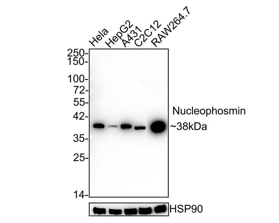
Western blot analysis of Nucleophosmin on different lysates with Rabbit anti-Nucleophosmin antibody (HA721206) at 1/1,000 dilution.
Lane 1: HeLa cell lysate
Lane 2: HepG2 cell lysate
Lane 3: A431 cell lysate
Lane 4: C2C12 cell lysate
Lane 5: RAW264.7 cell lysate
Lysates/proteins at 30 µg/Lane.
Predicted band size: 33 kDa
Observed band size: 38 kDa
Exposure time: 2 minutes;
4-20% SDS-PAGE gel.
Proteins were transferred to a PVDF membrane and blocked with 5% NFDM/TBST for 1 hour at room temperature. The primary antibody (HA721206) at 1/1,000 dilution was used in 5% NFDM/TBST at room temperature for 2 hours. Goat Anti-Rabbit IgG - HRP Secondary Antibody (HA1001) at 1/50,000 dilution was used for 1 hour at room temperature.
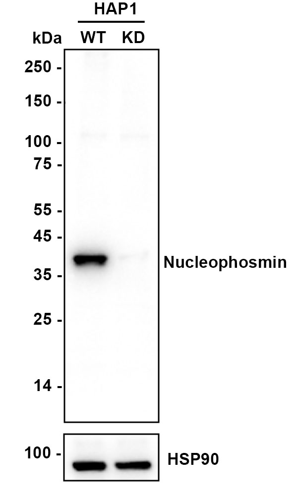
☑ Knockdown (KD)
Western blot analysis of Nucleophosmin on different lysates with Rabbit anti-Nucleophosmin antibody (HA721206) at 1/2,000 dilution.
Lane 1: HAP1-parental cell lysate
Lane 2: HAP1-Nucleophosmin KD cell lysate
Lysates/proteins at 10 µg/Lane.
Predicted band size: 33 kDa
Observed band size: 38 kDa
Exposure time: 4 seconds; ECL: K1801;
4-20% SDS-PAGE gel.
Proteins were transferred to a PVDF membrane and blocked with 5% NFDM/TBST for 1 hour at room temperature. The primary antibody (HA721206) at 1/2,000 dilution was used in K1803 at 4℃ overnight. Goat Anti-Rabbit IgG - HRP Secondary Antibody (HA1001) at 1/50,000 dilution was used for 1 hour at room temperature.
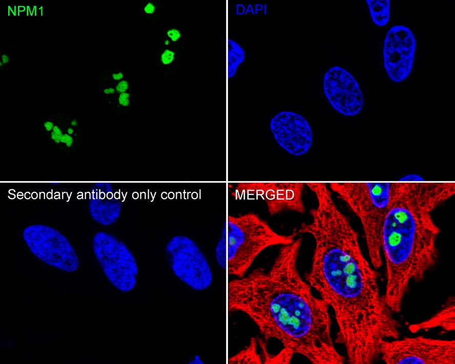
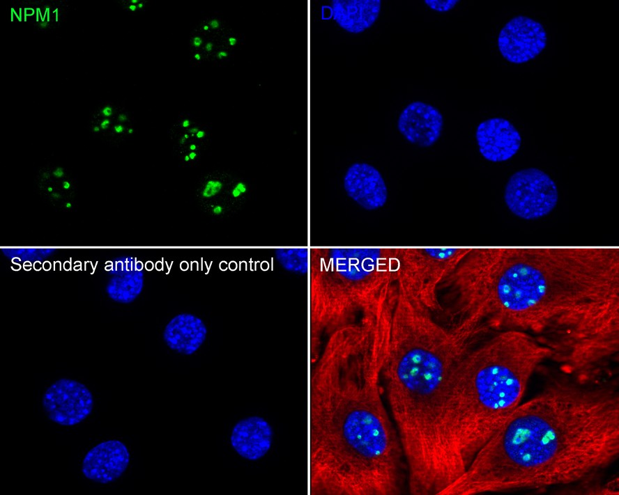
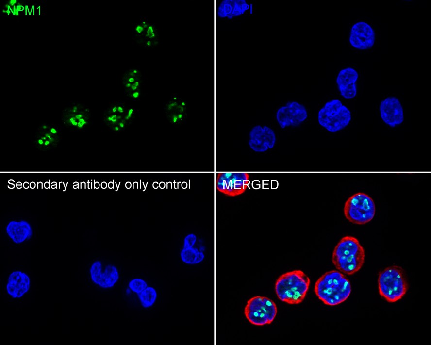
Immunocytochemistry analysis of HeLa cells labeling Nucleophosmin with Rabbit anti-Nucleophosmin antibody (HA721206) at 1/5,000 dilution.
Cells were fixed in 4% paraformaldehyde for 15 minutes at room temperature, permeabilized with 0.1% Triton X-100 in PBS for 15 minutes at room temperature, then blocked with 1% BSA in 10% negative goat serum for 1 hour at room temperature. Cells were then incubated with Rabbit anti-Nucleophosmin antibody (HA721206) at 1/5,000 dilution in 1% BSA in PBST overnight at 4 ℃. Goat Anti-Rabbit IgG H&L (iFluor™ 488, HA1121) was used as the secondary antibody at 1/1,000 dilution. PBS instead of the primary antibody was used as the secondary antibody only control. Nuclear DNA was labelled in blue with DAPI.
Beta tubulin (HA601187, red) was stained at 1/100 dilution overnight at +4℃. Goat Anti-Mouse IgG H&L (iFluor™ 594, HA1126) was used as the secondary antibody at 1/1,000 dilution.
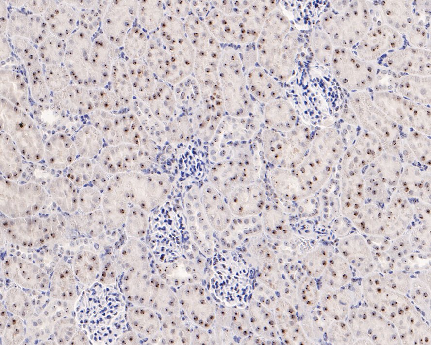
Immunohistochemical analysis of paraffin-embedded rat kidney tissue with Rabbit anti-Nucleophosmin antibody (HA721206) at 1/500 dilution.
The section was pre-treated using heat mediated antigen retrieval with sodium citrate buffer (pH 6.0) for 2 minutes. The tissues were blocked in 1% BSA for 20 minutes at room temperature, washed with ddH2O and PBS, and then probed with the primary antibody (HA721206) at 1/500 dilution for 1 hour at room temperature. The detection was performed using an HRP conjugated compact polymer system. DAB was used as the chromogen. Tissues were counterstained with hematoxylin and mounted with DPX.
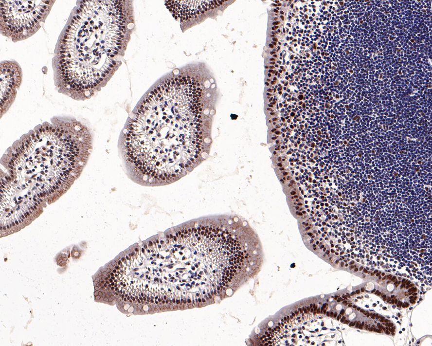
Immunohistochemical analysis of paraffin-embedded mouse small intestine tissue with Rabbit anti-Nucleophosmin antibody (HA721206) at 1/200 dilution.
The section was pre-treated using heat mediated antigen retrieval with sodium citrate buffer (pH 6.0) for 2 minutes. The tissues were blocked in 1% BSA for 20 minutes at room temperature, washed with ddH2O and PBS, and then probed with the primary antibody (HA721206) at 1/200 dilution for 1 hour at room temperature. The detection was performed using an HRP conjugated compact polymer system. DAB was used as the chromogen. Tissues were counterstained with hematoxylin and mounted with DPX.
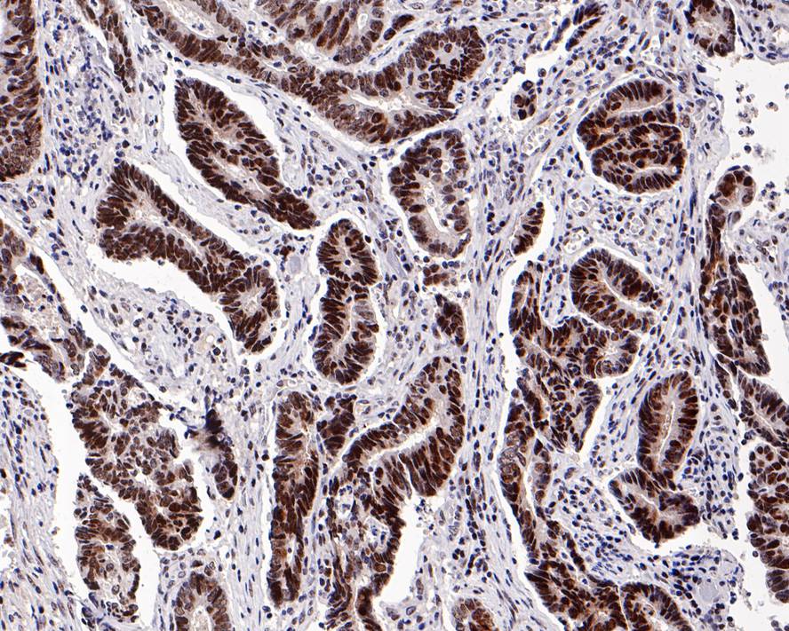
Immunohistochemical analysis of paraffin-embedded human colon carcinoma tissue with Rabbit anti-Nucleophosmin antibody (HA721206) at 1/200 dilution.
The section was pre-treated using heat mediated antigen retrieval with sodium citrate buffer (pH 6.0) for 2 minutes. The tissues were blocked in 1% BSA for 20 minutes at room temperature, washed with ddH2O and PBS, and then probed with the primary antibody (HA721206) at 1/200 dilution for 1 hour at room temperature. The detection was performed using an HRP conjugated compact polymer system. DAB was used as the chromogen. Tissues were counterstained with hematoxylin and mounted with DPX.
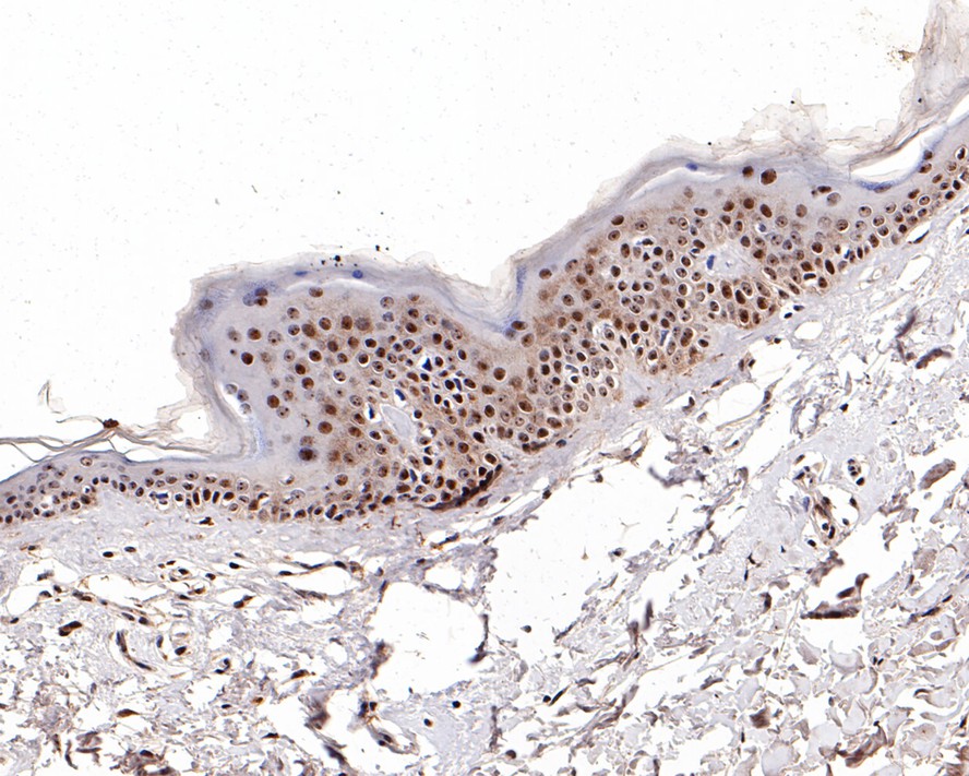
Immunohistochemical analysis of paraffin-embedded human skin tissue with Rabbit anti-Nucleophosmin antibody (HA721206) at 1/200 dilution.
The section was pre-treated using heat mediated antigen retrieval with sodium citrate buffer (pH 6.0) for 2 minutes. The tissues were blocked in 1% BSA for 20 minutes at room temperature, washed with ddH2O and PBS, and then probed with the primary antibody (HA721206) at 1/200 dilution for 1 hour at room temperature. The detection was performed using an HRP conjugated compact polymer system. DAB was used as the chromogen. Tissues were counterstained with hematoxylin and mounted with DPX.
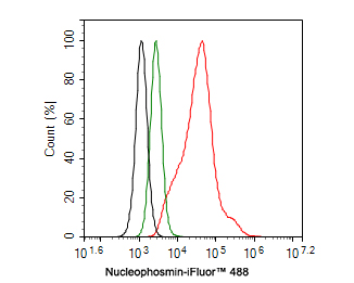
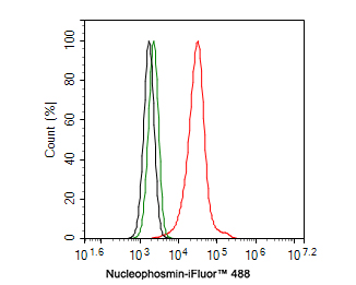
Flow cytometric analysis of Daudi cells labeling Nucleophosmin.
Cells were fixed and permeabilized. Then stained with the primary antibody (HA721206, 1ug/ml) (red) compared with Rabbit IgG Isotype Control (green). After incubation of the primary antibody at +4℃ for an hour, the cells were stained with a iFluor™ 488 conjugate-Goat anti-Rabbit IgG Secondary antibody (HA1121) at 1/1,000 dilution for 30 minutes at +4℃. Unlabelled sample was used as a control (cells without incubation with primary antibody; black).
Flow cytometric analysis of HL-60 cells labeling Nucleophosmin.
Cells were fixed and permeabilized. Then stained with the primary antibody (HA721206, 1ug/ml) (red) compared with Rabbit IgG Isotype Control (green). After incubation of the primary antibody at +4℃ for an hour, the cells were stained with a iFluor™ 488 conjugate-Goat anti-Rabbit IgG Secondary antibody (HA1121) at 1/1,000 dilution for 30 minutes at +4℃. Unlabelled sample was used as a control (cells without incubation with primary antibody; black).


Copyright © 广州杰特伟生物科技有限公司 All Rights Reserved. 备案号:粤ICP备19077843号