


LRP1 Recombinant Rabbit Monoclonal Antibody [SA0290]
Recombinant Rabbit monoclonal Antibody
Synthetic peptide within Human LRP1 aa 4,471-4,520 / 4,544.
Human, Mouse, Rat
WB, IF-Tissue, IHC-P, IP, IHC-Fr
Predicted band size: 505 kDa
Mouse liver tissue lysate, Human lung tissue lysate, Rat liver tissue lysate, Rat lung tissue lysate, Mouse lung tissue lysate, Human liver tissue lysate, mouse brain tissue, rat brain tissue, rat liver tissue, human lung tissue, human liver tissue, mouse liver tissue.
unconjugated
SA0290
Liquid
1ug/ul
Store at +4℃ after thawing. Aliquot store at -20℃ or -80℃. Avoid repeated freeze / thaw cycles.
1*TBS (pH7.4), 0.05% BSA, 40% Glycerol. Preservative: 0.05% Sodium Azide.
IgG
Protein A affinity purified.
WB
1:1,000-1:5,000
IF-Tissue
1:50
IHC-P
1:200-1:2,000
IP
1-2μg/sample
IHC-Fr
1:100
| Rat | 查看 1 篇文献如下 |
| Mouse | 查看 1 篇文献如下 |
LRP1 is a member of the LDLR family and ubiquitously expressed in multiple tissues, though it is most abundant in vascular smooth muscle cells (SMCs), hepatocytes, and neurons. LRP1 plays a key role in intracellular signaling and endocytosis, which thus implicate it in many cellular and biological processes, including lipid and lipoprotein metabolism, protease degradation, platelet derived growth factor receptor regulation, integrin maturation and recycling, regulation of vascular tone, regulation of blood brain barrier permeability, cell growth, cell migration, inflammation, and apoptosis, as well as diseases such as neurodegenerative diseases, atherosclerosis, and cancer. The LRP1 gene encodes a 600 kDa precursor protein that is processed by furin in the trans-Golgi complex, resulting in a 515 kDa alpha-chain and an 85 kDa beta-chain associated noncovalently.
1. Fernandez-Castaneda, A. et al. 2013. Identification of the low density lipoprotein (LDL) receptor-related protein-1 interactome in central nervous system myelin suggests a role in the clearance of necrotic cell debris. J. Biol. Chem. 288: 4538-4548.
2. Yahiro, K. et al. 2012. Low-density lipoprotein receptor-related protein-1 (LRP1) mediates autophagy and apoptosis caused by Helicobacter pylori VacA. J. Biol. Chem. 287: 31104-31115.
Belongs to the LDLR family.
Most abundant in liver, brain and lung.
Cleaved into a 85 kDa membrane-spanning subunit (LRP-85) and a 515 kDa large extracellular domain (LRP-515) that remains non-covalently associated. Gamma-secretase-dependent cleavage of LRP-85 releases the intracellular domain from the membrane.; The N-terminus is blocked.; Phosphorylated on serine and threonine residues.; Phosphorylated on tyrosine residues upon stimulation with PDGF. Tyrosine phosphorylation promotes interaction with SHC1.
Cytoplasm, Nucleus, Membrane.
A2MR antibody
Alpha 2 macroglobulin receptor antibody
alpha 2MR antibody
Alpha-2-macroglobulin receptor antibody
APOER antibody
Apolipoprotein E receptor antibody
APR antibody
CD 91 antibody
CD91 antibody
CD91 antigen antibody
展开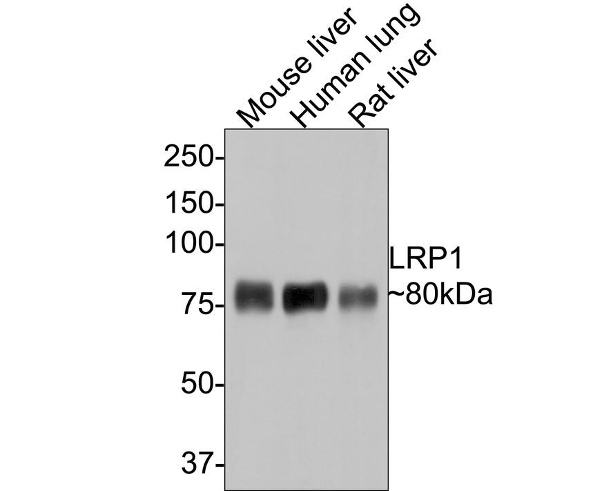
Western blot analysis of LRP1 on different lysates with Rabbit anti-LRP1 antibody (ET1601-1) at 1/5,000 dilution.
Lane 1: Mouse liver tissue lysate
Lane 2: Human lung tissue lysate
Lane 3: Rat liver tissue lysate
Lysates/proteins at 20 µg/Lane.
Predicted band size: 505 kDa
Observed band size: 80 kDa
Exposure time: 30 seconds;
8% SDS-PAGE gel.
Proteins were transferred to a PVDF membrane and blocked with 5% NFDM/TBST for 1 hour at room temperature. The primary antibody (ET1601-1) at 1/5,000 dilution was used in 5% NFDM/TBST at room temperature for 2 hours. Goat Anti-Rabbit IgG - HRP Secondary Antibody (HA1001) at 1:300,000 dilution was used for 1 hour at room temperature.
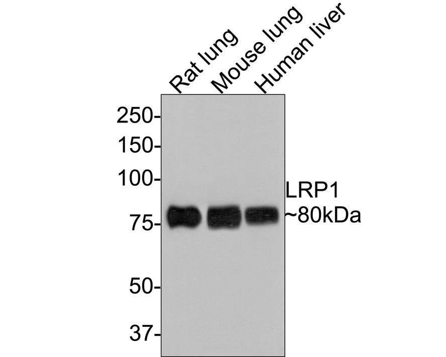
Western blot analysis of LRP1 on different lysates with Rabbit anti-LRP1 antibody (ET1601-1) at 1/5,000 dilution.
Lane 1: Rat lung tissue lysate
Lane 2: Mouse lung tissue lysate
Lane 3: Human liver tissue lysate
Lysates/proteins at 20 µg/Lane.
Predicted band size: 505 kDa
Observed band size: 80 kDa
Exposure time: 1 minute;
8% SDS-PAGE gel.
Proteins were transferred to a PVDF membrane and blocked with 5% NFDM/TBST for 1 hour at room temperature. The primary antibody (ET1601-1) at 1/5,000 dilution was used in 5% NFDM/TBST at room temperature for 2 hours. Goat Anti-Rabbit IgG - HRP Secondary Antibody (HA1001) at 1:300,000 dilution was used for 1 hour at room temperature.
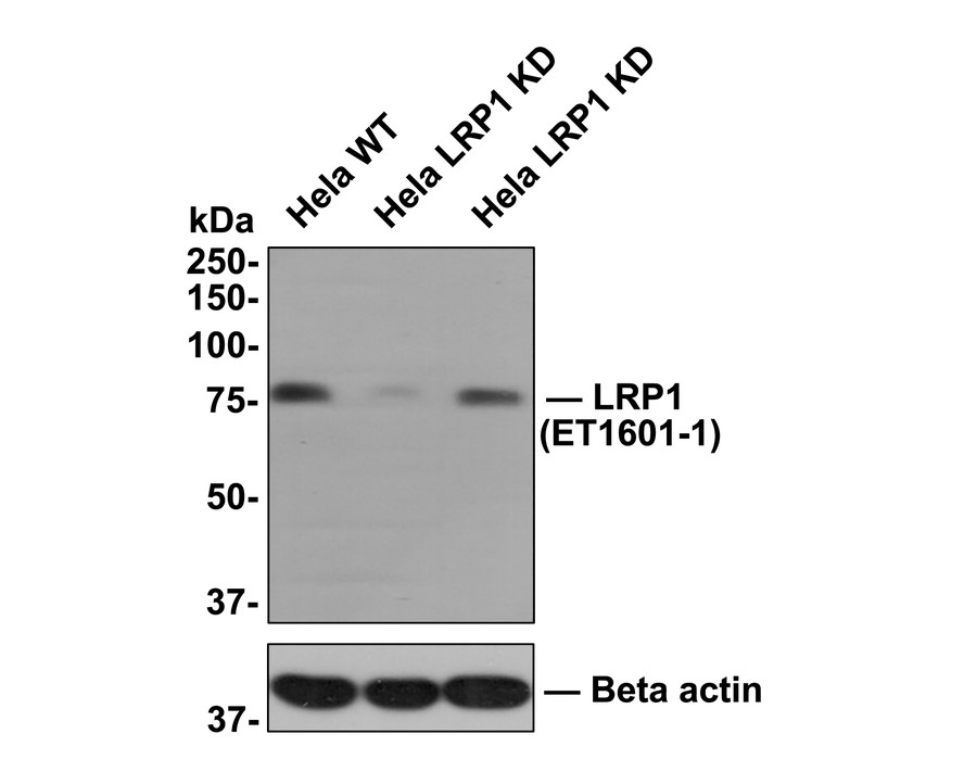
☑ Knockdown (KD)
All lanes: Western blot analysis of LRP1 with anti-LRP1 antibody (ET1601-1) at 1:1,000 dilution.
Lane 1: Wild-type Hela whole cell lysate (10 µg).
Lane 2/3: LRP1 knockdown Hela whole cell lysate (10 µg).
ET1601-1 was shown to specifically react with LRP1 in wild-type Hela cells. Weakened bands were observed when LRP1 knockdown samples were tested. Wild-type and LRP1 knockdown samples were subjected to SDS-PAGE. Proteins were transferred to a PVDF membrane and blocked with 5% NFDM in TBST for 1 hour at room temperature. The primary antibody (ET1601-1, 1/1,000) was used in 5% BSA at room temperature for 2 hours. Goat Anti-Rabbit IgG-HRP Secondary Antibody (HA1001) at 1:300,000 dilution was used for 1 hour at room temperature.
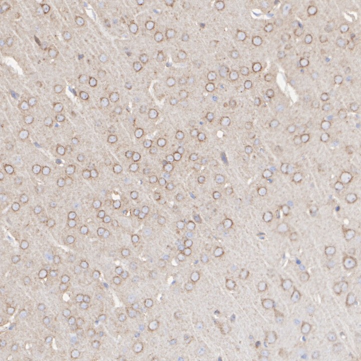
Immunohistochemical analysis of paraffin-embedded mouse brain tissue with Rabbit anti-LRP1 antibody (ET1601-1) at 1/2,000 dilution.
The section was pre-treated using heat mediated antigen retrieval with sodium citrate buffer (pH 6.0) for 2 minutes. The tissues were blocked in 1% BSA for 20 minutes at room temperature, washed with ddH2O and PBS, and then probed with the primary antibody (ET1601-1) at 1/2,000 dilution for 1 hour at room temperature. The detection was performed using an HRP conjugated compact polymer system. DAB was used as the chromogen. Tissues were counterstained with hematoxylin and mounted with DPX.
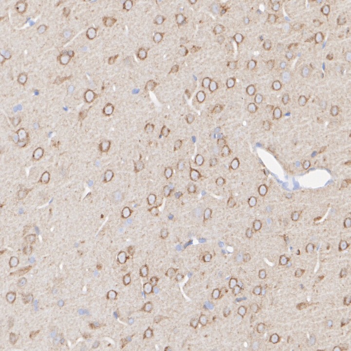
Immunohistochemical analysis of paraffin-embedded rat brain tissue with Rabbit anti-LRP1 antibody (ET1601-1) at 1/2,000 dilution.
The section was pre-treated using heat mediated antigen retrieval with sodium citrate buffer (pH 6.0) for 2 minutes. The tissues were blocked in 1% BSA for 20 minutes at room temperature, washed with ddH2O and PBS, and then probed with the primary antibody (ET1601-1) at 1/2,000 dilution for 1 hour at room temperature. The detection was performed using an HRP conjugated compact polymer system. DAB was used as the chromogen. Tissues were counterstained with hematoxylin and mounted with DPX.
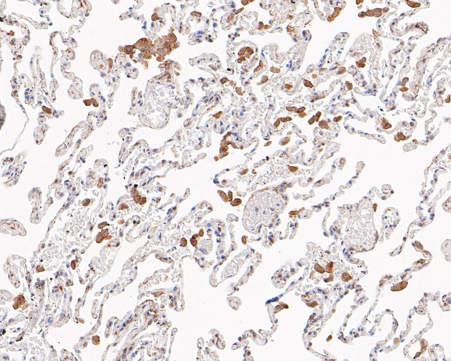
Immunohistochemical analysis of paraffin-embedded human lung tissue with Rabbit anti-LRP1 antibody (ET1601-1) at 1/200 dilution.
The section was pre-treated using heat mediated antigen retrieval with sodium citrate buffer (pH 6.0) for 2 minutes. The tissues were blocked in 1% BSA for 20 minutes at room temperature, washed with ddH2O and PBS, and then probed with the primary antibody (ET1601-1) at 1/200 dilution for 1 hour at room temperature. The detection was performed using an HRP conjugated compact polymer system. DAB was used as the chromogen. Tissues were counterstained with hematoxylin and mounted with DPX.

Immunohistochemical analysis of paraffin-embedded human liver tissue with Rabbit anti-LRP1 antibody (ET1601-1) at 1/2,000 dilution.
The section was pre-treated using heat mediated antigen retrieval with sodium citrate buffer (pH 6.0) for 2 minutes. The tissues were blocked in 1% BSA for 20 minutes at room temperature, washed with ddH2O and PBS, and then probed with the primary antibody (ET1601-1) at 1/2,000 dilution for 1 hour at room temperature. The detection was performed using an HRP conjugated compact polymer system. DAB was used as the chromogen. Tissues were counterstained with hematoxylin and mounted with DPX.
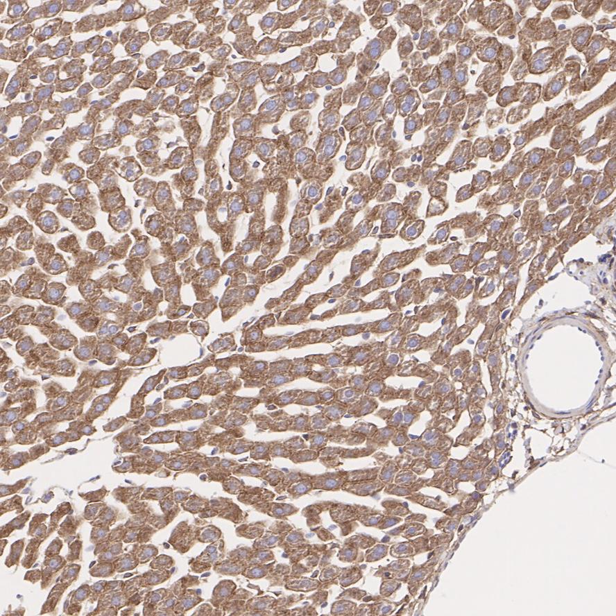
Immunohistochemical analysis of paraffin-embedded mouse liver tissue with Rabbit anti-LRP1 antibody (ET1601-1) at 1/200 dilution.
The section was pre-treated using heat mediated antigen retrieval with sodium citrate buffer (pH 6.0) for 2 minutes. The tissues were blocked in 1% BSA for 20 minutes at room temperature, washed with ddH2O and PBS, and then probed with the primary antibody (ET1601-1) at 1/200 dilution for 1 hour at room temperature. The detection was performed using an HRP conjugated compact polymer system. DAB was used as the chromogen. Tissues were counterstained with hematoxylin and mounted with DPX.

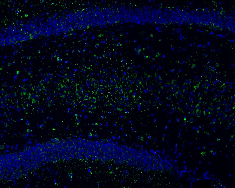
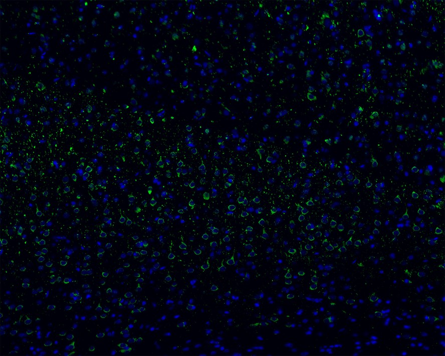
Immunofluorescence analysis of frozen mouse cerebral cortex tissue labeling LRP1 with Rabbit anti-LRP1 antibody (ET1601-1).
The tissues were blocked in 3% BSA for 30 minutes at room temperature, washed with PBS, and then probed with the primary antibody (ET1601-1, green) at 1/100 dilution overnight at 4℃, washed with PBS. Goat Anti-Rabbit IgG H&L (Alexa Fluor® 488) was used as the secondary antibody at 1/200 dilution. Nuclei were counterstained with DAPI (blue). Image acquisition was performed with KFBIO KF-FL-400 Scanner.
Schisandra chinensis lignans exerts endocannabinoids-like antidepressive effect: The phagocytotic relationship of activated CB2R-mediated M2 microglia and “stressed-but-viable” neuron
Author: Jinyu Wang, Haoyu Du, Mengru Li, Tingxu Yan, Ying Jia
PMID: 39832627
期刊: Journal Of Ethnopharmacology
应用: WB
反应种属: Rat,Mouse
发表时间: 2025 Jan


Copyright © 广州杰特伟生物科技有限公司 All Rights Reserved. 备案号:粤ICP备19077843号