


Bcl-2 Recombinant Rabbit Monoclonal Antibody [PD01-41]
Recombinant Rabbit monoclonal Antibody
Synthetic peptide within Human Bcl2 alpha aa 50-150 (N terminal).
Human
WB, IHC-P, IF-Cell, FC
Predicted band size: 26 kDa
MCF7 cell lysate, Jurkat cell lysate, HL-60 cell lysate, THP-1 cell lysate, human B-cell lymphoma tissue, human tonsil tissue, human lymph nodes tissue, HeLa, THP-1.
unconjugated
PD01-41
Liquid
1ug/ul
Store at +4℃ after thawing. Aliquot store at -20℃. Avoid repeated freeze / thaw cycles.
PBS (pH7.4), 0.1% BSA, 40% Glycerol. Preservative: 0.05% Sodium Azide.
IgG
Protein A affinity purified.
WB
1:2,000-1:5,000
IHC-P
1:1,000-1:5,000
IF-Cell
1:100
FC
1:1,000
| WB | 查看 6 篇文献如下 |
| IHC | 查看 1 篇文献如下 |
| Human | 查看 4 篇文献如下 |
| rat | 查看 1 篇文献如下 |
| Rat | 查看 1 篇文献如下 |
Proteins of the Bcl-2 family are regulators of apoptosis (programmed cell death) localized to membranes of primarily mitochondria, but also to smooth endoplasmic reticulum and nucleolemma. The fine balance between pro- and anti-apoptotic Bcl-2 family members regulates the cell fate in response to many signalling pathways. Altered expression of the proteins may lead to either premature cell death or to inappropriate cell survival promoting neoplastic growth. Bcl-2, Bax and Bcl-X are the most well known proteins in this family. In neoplastic lesions Bcl-2 upregulation may act by suppression of programmed cell death and extension of the tumour cell life span. Bcl-2 overexpression contributes to increased resistance to chemotherapy. However, the prognostic value of Bcl-2 overexpression is dependent on the tumour type, and in some types BCl-2 may even have a tumour suppressor effect. Overexpression of Bcl-2 is common in many types of cancer, including non-Hodgkin's lymphoma and leukaemias, adenocarcinomas (e.g., prostate, colorectum, stomach, and lung), squamous cell carcinoma, small cell carcinoma, neuroblastoma and various sarcomas. Bcl-2 is helpful in distinguishing follicular lymphoma from reactive follicular hyperplasia. Bcl-2 staining can not be used for differential diagnosis between follicular lymphoma and other types of low-grade B-cell lymphomas since most of the latter also express bcl-2. Monocytoid B-cell hyperplasia contrasts with bcl-2 negativity against positive reaction in 80% of the marginal zone lymphomas. Possibly, Bcl-2 may aid in the differential diagnosis of sarcomas. Tonsil is recommendable as positive and negative tissue control for BCL2. A moderate to strong, predominantly cytoplasmic staining reaction should be displayed in virtually all T-cells and B-cells in the mantle zone of the reactive follicles, whereas the majority of the basal squamous epithelial cells, e.g. lining the tonsillar crypts, must show an at least weak staining intensity. Germinal centre T-cells should be distinctively demonstrated, whereas germinal centre B-cells should be negative.
1. Cao LH et al. Morphine, a potential antagonist of cisplatin cytotoxicity, inhibits cisplatin-induced apoptosis and suppression of tumor growth in nasopharyngeal carcinoma xenografts. Sci Rep 6:18706 (2016).
2. Chen B et al. Inhibition of miR-29c promotes proliferation, and inhibits apoptosis and differentiation in P19 embryonic carcinoma cells. Mol Med Rep 13:2527-35 (2016).
Belongs to the Bcl-2 family.
Expressed in a variety of tissues.
Phosphorylation/dephosphorylation on Ser-70 regulates anti-apoptotic activity. Growth factor-stimulated phosphorylation on Ser-70 by PKC is required for the anti-apoptosis activity and occurs during the G2/M phase of the cell cycle. In the absence of growth factors, BCL2 appears to be phosphorylated by other protein kinases such as ERKs and stress-activated kinases. Phosphorylated by MAPK8/JNK1 at Thr-69, Ser-70 and Ser-87, wich stimulates starvation-induced autophagy. Dephosphorylated by protein phosphatase 2A (PP2A) (By similarity).; Proteolytically cleaved by caspases during apoptosis. The cleaved protein, lacking the BH4 motif, has pro-apoptotic activity, causes the release of cytochrome c into the cytosol promoting further caspase activity.; Monoubiquitinated by PRKN, leading to increase its stability. Ubiquitinated by SCF(FBXO10), leading to its degradation by the proteasome.
Mitochondrion outer membrane, Nucleus membrane, Endoplasmic reticulum membrane, Cytoplasm.
Apoptosis regulator Bcl 2 antibody
Apoptosis regulator Bcl-2 antibody
Apoptosis regulator Bcl2 antibody
AW986256 antibody
B cell CLL/lymphoma 2 antibody
B cell leukemia/lymphoma 2 antibody
Bcl-2 antibody
Bcl2 antibody
BCL2_HUMAN antibody
C430015F12Rik antibody
展开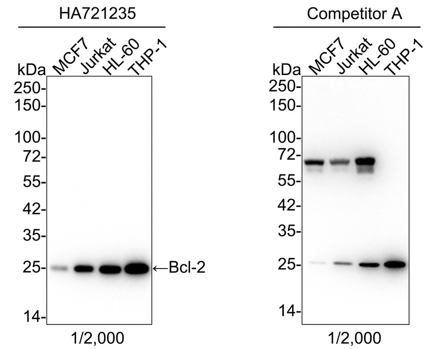
Western blot analysis of Bcl-2 on different lysates with Rabbit anti-Bcl-2 antibody (HA721235) at 1/2,000 dilution and competitor's antibody at 1/2,000 dilution.
Lane 1: MCF7 cell lysate (15 µg/Lane)
Lane 2: Jurkat cell lysate (15 µg/Lane)
Lane 3: HL-60 cell lysate (15 µg/Lane)
Lane 4: THP-1 cell lysate (15 µg/Lane)
Predicted band size: 26 kDa
Observed band size: 25 kDa
Exposure time: 1 minute 30 seconds; ECL: K1801;
4-20% SDS-PAGE gel.
Proteins were transferred to a PVDF membrane and blocked with 5% NFDM/TBST for 1 hour at room temperature. The primary antibody (HA721235) at 1/2,000 dilution and competitor's antibody at 1/2,000 dilution were used in 5% NFDM/TBST at 4℃ overnight. Goat Anti-Rabbit IgG - HRP Secondary Antibody (HA1001) at 1/50,000 dilution was used for 1 hour at room temperature.
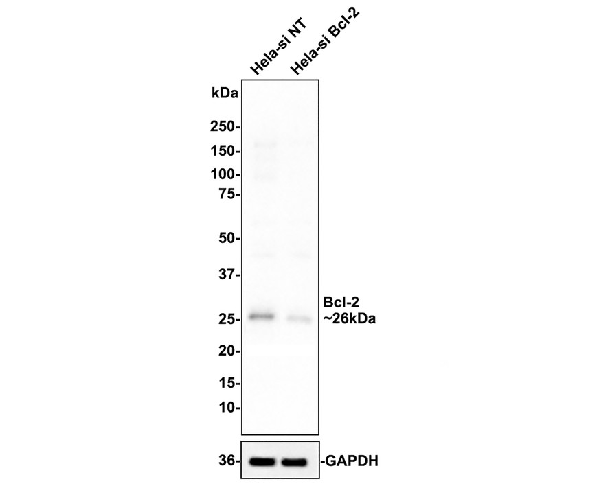
☑ Knockdown (KD)
Western blot analysis of Bcl-2 on different lysates with Rabbit anti-Bcl-2 antibody (HA721235) at 1/1,000 dilution.
Lane 1: Hela-si NT cell lysate (10 µg/Lane)
Lane 2: Hela-si Bcl-2 cell lysate (10 µg/Lane)
Predicted band size: 26 kDa
Observed band size: 26 kDa
Exposure time: 31 seconds;
ECL: merk
4-20% SDS-PAGE gel.
HA721235 was shown to specifically react with Bcl-2 in Hela-si NT cells. Weakened band was observed when Hela-si Bcl-2 sample was tested. Hela-si NT and Hela-si Bcl-2 samples were subjected to SDS-PAGE. Proteins were transferred to a PVDF membrane and blocked with 5% NFDM in TBST for 1 hour at room temperature. The primary antibody (HA721235, 1/1,000) and Loading control antibody (Rabbit anti-GAPDH, ET1601-4, 1/10,000) were used in 5% BSA at room temperature for 2 hours. Goat Anti-rabbit IgG-HRP Secondary Antibody (HA1001) at 1:100,000 dilution was used for 1 hour at room temperature.
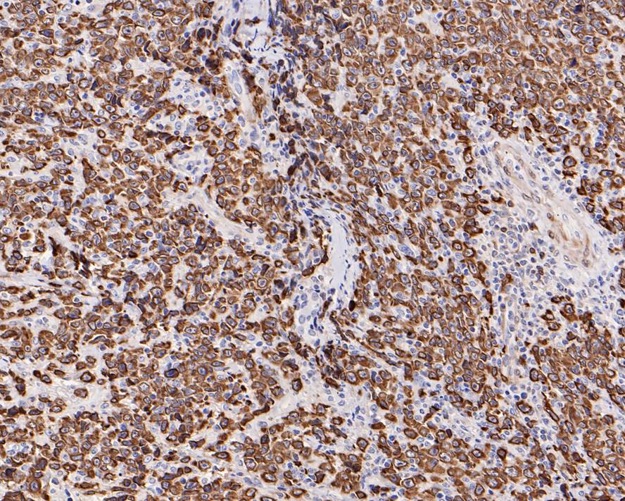
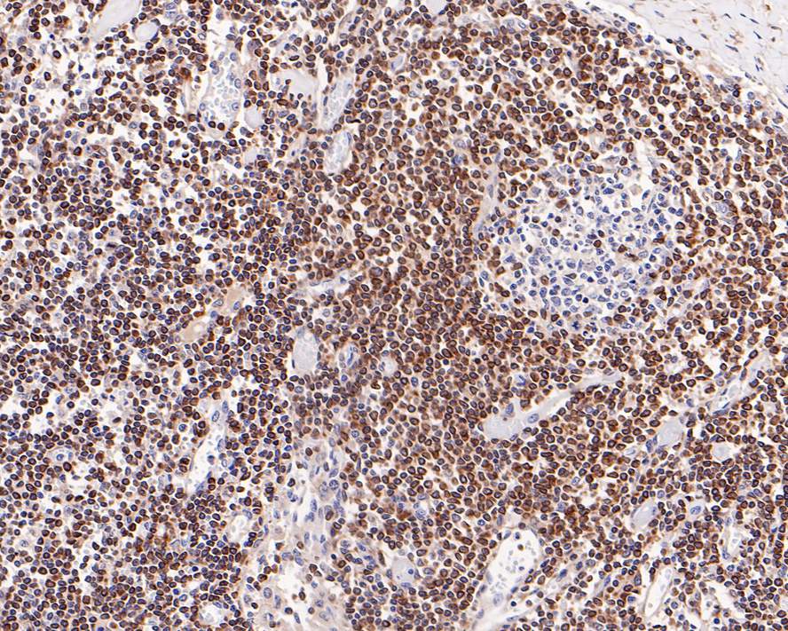
Immunohistochemical analysis of paraffin-embedded human B-cell lymphoma tissue with Rabbit anti-Bcl-2 antibody (HA721235) at 1/1,000 dilution.
The section was pre-treated using heat mediated antigen retrieval with Tris-EDTA buffer (pH 9.0) for 20 minutes. The tissues were blocked in 1% BSA for 20 minutes at room temperature, washed with ddH2O and PBS, and then probed with the primary antibody (HA721235) at 1/1,000 dilution for 1 hour at room temperature. The detection was performed using an HRP conjugated compact polymer system. DAB was used as the chromogen. Tissues were counterstained with hematoxylin and mounted with DPX.
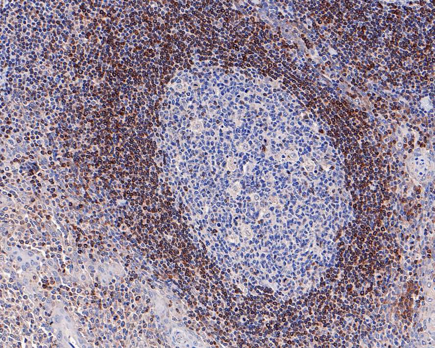
Immunohistochemical analysis of paraffin-embedded human tonsil tissue with Rabbit anti-Bcl-2 antibody (HA721235) at 1/2,000 dilution.
The section was pre-treated using heat mediated antigen retrieval with Tris-EDTA buffer (pH 9.0) for 20 minutes. The tissues were blocked in 1% BSA for 20 minutes at room temperature, washed with ddH2O and PBS, and then probed with the primary antibody (HA721235) at 1/2,000 dilution for 1 hour at room temperature. The detection was performed using an HRP conjugated compact polymer system. DAB was used as the chromogen. Tissues were counterstained with hematoxylin and mounted with DPX.
Immunohistochemical analysis of paraffin-embedded human lymph nodes tissue with Rabbit anti-Bcl-2 antibody (HA721235) at 1/5,000 dilution.
The section was pre-treated using heat mediated antigen retrieval with Tris-EDTA buffer (pH 9.0) for 20 minutes. The tissues were blocked in 1% BSA for 20 minutes at room temperature, washed with ddH2O and PBS, and then probed with the primary antibody (HA721235) at 1/5,000 dilution for 1 hour at room temperature. The detection was performed using an HRP conjugated compact polymer system. DAB was used as the chromogen. Tissues were counterstained with hematoxylin and mounted with DPX.

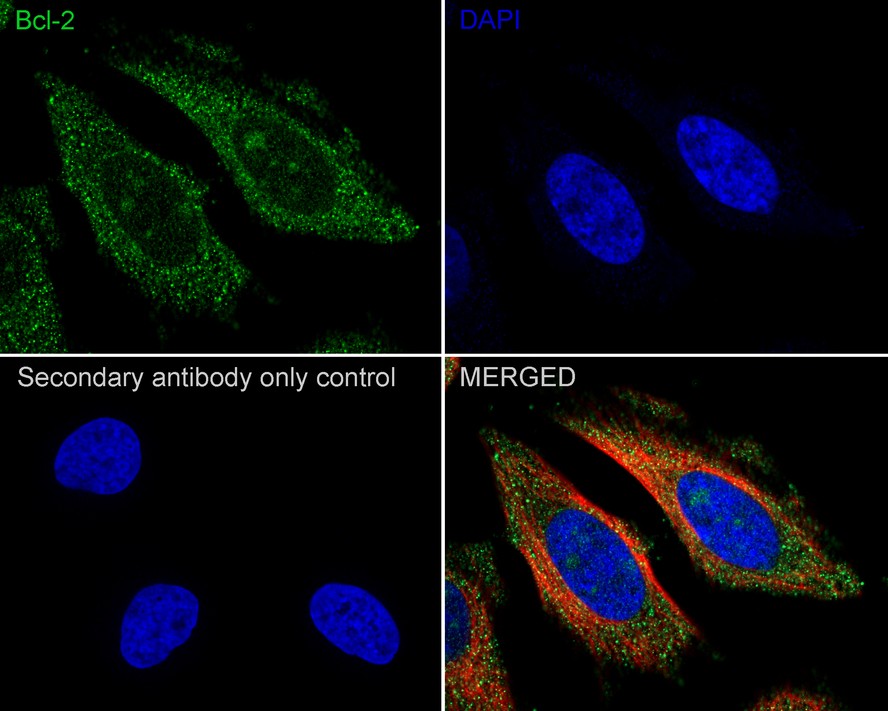
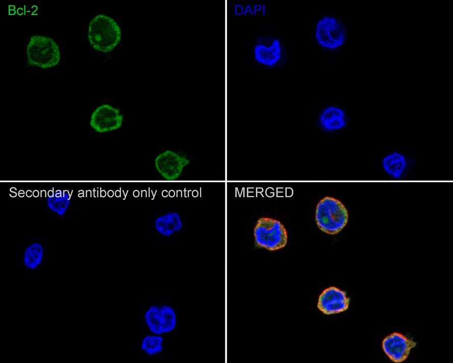
Immunocytochemistry analysis of HeLa cells labeling Bcl-2 with Rabbit anti-Bcl-2 antibody (HA721235) at 1/100 dilution.
Cells were fixed in 4% paraformaldehyde for 20 minutes at room temperature, permeabilized with 0.1% Triton X-100 in PBS for 5 minutes at room temperature, then blocked with 1% BSA in 10% negative goat serum for 1 hour at room temperature. Cells were then incubated with Rabbit anti-Bcl-2 antibody (HA721235) at 1/100 dilution in 1% BSA in PBST overnight at 4 ℃. Goat Anti-Rabbit IgG H&L (iFluor™ 488, HA1121) was used as the secondary antibody at 1/1,000 dilution. PBS instead of the primary antibody was used as the secondary antibody only control. Nuclear DNA was labelled in blue with DAPI.
Beta tubulin (M1305-2, red) was stained at 1/100 dilution overnight at +4℃. Goat Anti-Mouse IgG H&L (iFluor™ 594, HA1126) was used as the secondary antibody at 1/1,000 dilution.
Immunocytochemistry analysis of THP-1 cells labeling Bcl-2 with Rabbit anti-Bcl-2 antibody (HA721235) at 1/100 dilution.
Cells were fixed in 4% paraformaldehyde for 20 minutes at room temperature, permeabilized with 0.1% Triton X-100 in PBS for 5 minutes at room temperature, then blocked with 1% BSA in 10% negative goat serum for 1 hour at room temperature. Cells were then incubated with Rabbit anti-Bcl-2 antibody (HA721235) at 1/100 dilution in 1% BSA in PBST overnight at 4 ℃. Goat Anti-Rabbit IgG H&L (iFluor™ 488, HA1121) was used as the secondary antibody at 1/1,000 dilution. PBS instead of the primary antibody was used as the secondary antibody only control. Nuclear DNA was labelled in blue with DAPI.
Beta tubulin (M1305-2, red) was stained at 1/100 dilution overnight at +4℃. Goat Anti-Mouse IgG H&L (iFluor™ 594, HA1126) was used as the secondary antibody at 1/1,000 dilution.

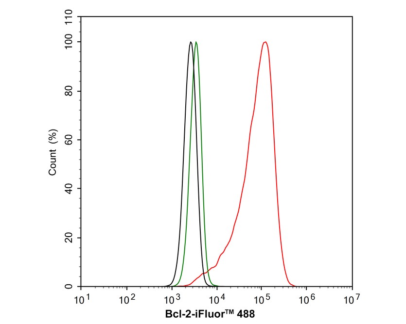
Flow cytometric analysis of HeLa cells labeling Bcl-2.
Cells were fixed and permeabilized. Then stained with the primary antibody (HA721235, 1μg/mL) (red) compared with Rabbit IgG Isotype Control (green). After incubation of the primary antibody at +4℃ for an hour, the cells were stained with a iFluor™ 488 conjugate-Goat anti-Rabbit IgG Secondary antibody (HA1121) at 1/1,000 dilution for 30 minutes at +4℃. Unlabelled sample was used as a control (cells without incubation with primary antibody; black).
Downregulation of miR-27a-3p induces endothelial injury and senescence and its significance in the development of coronary heart disease
Author: Chong-Yang Song, Hai-Zhen Huang, Ting-Ting Yan, Chen-Xi Cui, Hua-Yu Wu, Jing Chen, Jun-Hua Peng, Ning-Yuan Chen, Jun Tang, Shang-Ling Pan
PMID: 40147550
期刊: Cellular Signalling
应用: WB
反应种属: Human
发表时间: 2025 Mar
Citation


Copyright © 广州杰特伟生物科技有限公司 All Rights Reserved. 备案号:粤ICP备19077843号