


Oct4 Recombinant Rabbit Monoclonal Antibody [SD0750]
Recombinant Rabbit monoclonal Antibody
Synthetic peptide within human Oct4 aa 20-60.
Human, Mouse
WB, IF-Cell, IF-Tissue, IHC-P, IP, FC, ChIP
Predicted band size: 39 kDa
F9 cell lysate, NCCIT cell lysate, NCCIT, F9, mouse liver tissue, human seminoma tissue tissue.
unconjugated
SD0750
Liquid
1ug/ul
Store at +4℃ after thawing. Aliquot store at -20℃ or -80℃. Avoid repeated freeze / thaw cycles.
1*TBS (pH7.4), 0.05% BSA, 40% Glycerol. Preservative: 0.05% Sodium Azide.
IgG
Protein A affinity purified.
WB
1:2,000-1:8,000
IF-Cell
1:200-1:2,000
IF-Tissue
1:2,000
IHC-P
1:500-1:4,000
IP
Use at an assay dependent concentration.
FC
1ug/mL
ChIP
Use 0.5~2 μg for 25 μg of chromatin.
| Human | 查看 5 篇文献如下 |
| Mouse | 查看 2 篇文献如下 |
| Human | 查看 1 篇文献如下 |
POU5F1 (POU domain, class 5, transcription factor 1), also known as octamer-binding transcription factor-3 (Oct-3, OTF3), octamer-binding transcription factor-4 (Oct-4, Otf-4) and Oct-3/4, modulates embryonic stem (ES) cell populations by influencing lineage commitment. Oct-3/4 sustains stem-cell self-renewal and differentiation pathways. Transcription factors containing the POU homeodomain regulate tissue-specific gene expression in lymphoid and pituitary differentiation and in early mammalian development. Oct-3/4 is capable of inducing rapid proliferation and tumorigenic properties of ES cells through activation of the UTF1 gene. In humans, two Oct-3/4 isoforms contribute to influencing the undifferentiated phenotype of ES cells. Oct-3/4 pseudogenes localizing to human chromosomes 10 and 8 are reported to be transcribed in certain cancer cell lines and tissues.
1. Vessoni AT et al. Cockayne syndrome-derived neurons display reduced synapse density and altered neural network synchrony. Hum Mol Genet 25:1271-80 (2016).
2. Fang L et al. Jumonji AT-rich interactive domain 1B overexpression is associated with the development and progression of glioma. Int J Mol Med 38:172-82 (2016).
Belongs to the POU transcription factor family. Class-5 subfamily.
Expressed in developing brain. Highest levels found in specific cell layers of the cortex, the olfactory bulb, the hippocampus and the cerebellum. Low levels of expression in adult tissues.
Sumoylation enhances the protein stability, DNA binding and transactivation activity. Sumoylation is required for enhanced YES1 expression.; Ubiquitinated; undergoes 'Lys-63'-linked polyubiquitination by WWP2 leading to proteasomal degradation.; ERK1/2-mediated phosphorylation at Ser-111 promotes nuclear exclusion and proteasomal degradation. Phosphorylation at Thr-235 and Ser-236 decrease DNA-binding and alters ability to activate transcription.
Cytoplasm, Nucleus.
Octamer binding transcription factor 4 antibody
MGC22487 antibody
Oct 3 antibody
Oct 4 antibody
Oct-3 antibody
Oct-4 antibody
OCT3 antibody
Oct4 antibody
Octamer binding protein 3 antibody
Octamer binding protein 4 antibody
展开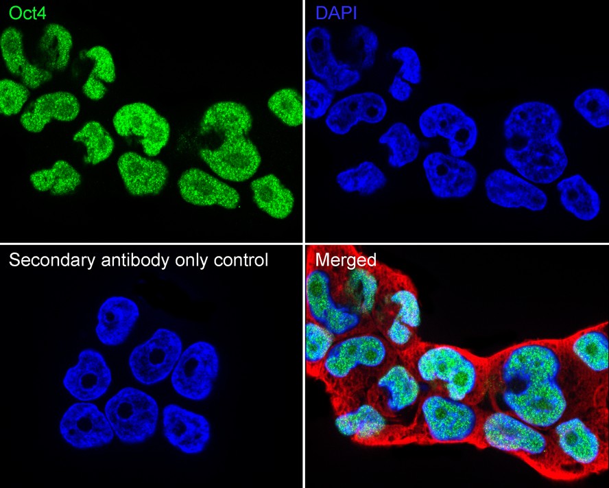
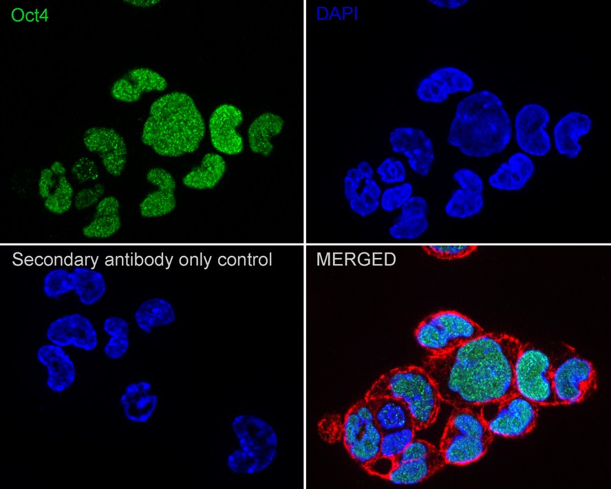
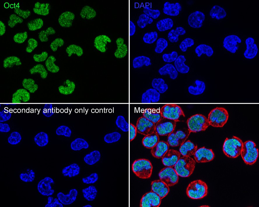
Immunocytochemistry analysis of NCCIT cells labeling Oct4 with Rabbit anti-Oct4 antibody (ET1612-20) at 1/200 dilution.
Cells were fixed in 4% paraformaldehyde for 20 minutes at room temperature, permeabilized with 0.1% Triton X-100 in PBS for 5 minutes at room temperature, then blocked with 1% BSA in 10% negative goat serum for 1 hour at room temperature. Cells were then incubated with Rabbit anti-Oct4 antibody (ET1612-20) at 1/200 dilution in 1% BSA in PBST overnight at 4 ℃. Goat Anti-Rabbit IgG H&L (iFluor™ 488, HA1121) was used as the secondary antibody at 1/1,000 dilution. PBS instead of the primary antibody was used as the secondary antibody only control. Nuclear DNA was labelled in blue with DAPI.
Beta tubulin (M1305-2, red) was stained at 1/100 dilution overnight at +4℃. Goat Anti-Mouse IgG H&L (iFluor™ 594, HA1126) was used as the secondary antibody at 1/1,000 dilution.
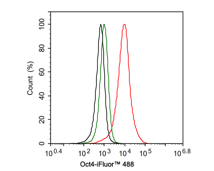
Flow cytometric analysis of NCCIT cells labeling Oct4.
Cells were fixed and permeabilized. Then stained with the primary antibody (ET1612-20, 1ug/ml) (red) compared with Rabbit IgG Isotype Control (green). After incubation of the primary antibody at +4℃ for an hour, the cells were stained with a iFluor™ 488 conjugate-Goat anti-Rabbit IgG Secondary antibody (HA1121) at 1/1,000 dilution for 30 minutes at +4℃. Unlabelled sample was used as a control (cells without incubation with primary antibody; black).

☑ Knockout (KO)
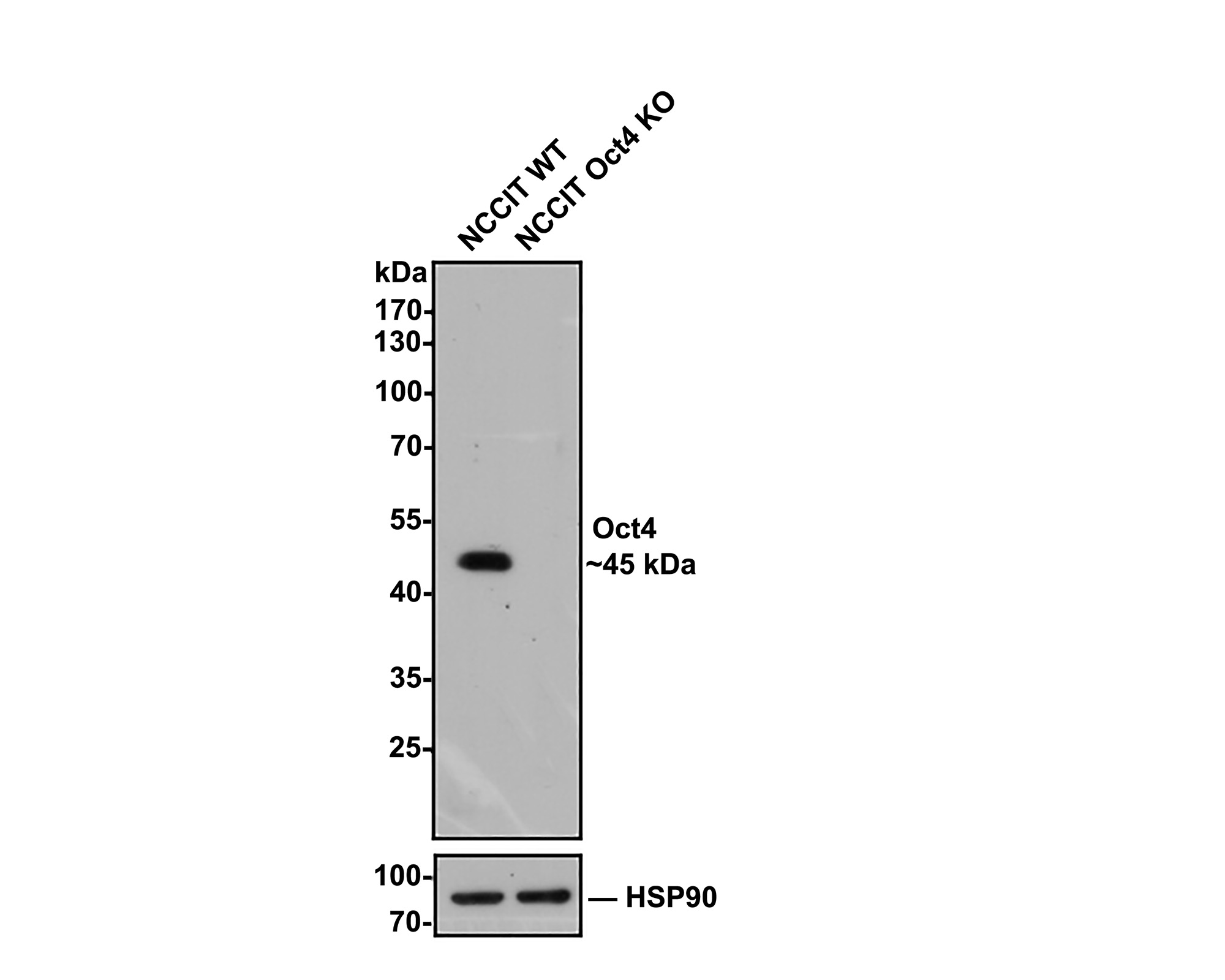
Western blot analysis of Oct4 with anti-Oct4 antibody (ET1612-20) at 1/2,000 dilution.
Lane 1: Wild-type NCCIT whole cell lysate.
Lane 2: Oct4 knockout NCCIT whole cell lysate.
Proteins were transferred to a PVDF membrane and blocked with 5% NFDM in TBST for 1 hour at room temperature. The primary Anti-Oct4 antibody (ET1612-20, 1/2,000) and Anti-HSP90 antibody (ET1605-56, 1/10,000) were used in 5% BSA at room temperature for 2 hours. Goat Anti-Rabbit IgG H&L (HRP) Secondary Antibody (HA1001) at 1/50,000 dilution was used for 1 hour at room temperature.

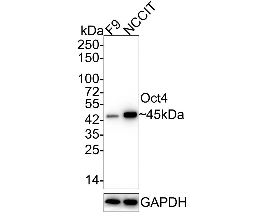
Western blot analysis of Oct4 on different lysates with Rabbit anti-Oct4 antibody (ET1612-20) at 1/2,000 dilution.
Lane 1: F9 cell lysate
Lane 2: NCCIT cell lysate
Lysates/proteins at 20 µg/Lane.
Predicted band size: 39 kDa
Observed band size: 45 kDa
Exposure time: 1 minute 59 seconds;
4-20% SDS-PAGE gel.
Proteins were transferred to a PVDF membrane and blocked with 5% NFDM/TBST for 1 hour at room temperature. The primary antibody (ET1612-20) at 1/2,000 dilution was used in 5% NFDM/TBST at 4℃ overnight. Goat Anti-Rabbit IgG - HRP Secondary Antibody (HA1001) at 1/50,000 dilution was used for 1 hour at room temperature.
Immunocytochemistry analysis of F9 cells labeling Oct4 with Rabbit anti-Oct4 antibody (ET1612-20) at 1/200 dilution.
Cells were fixed in 4% paraformaldehyde for 20 minutes at room temperature, permeabilized with 0.1% Triton X-100 in PBS for 5 minutes at room temperature, then blocked with 1% BSA in 10% negative goat serum for 1 hour at room temperature. Cells were then incubated with Rabbit anti-Oct4 antibody (ET1612-20) at 1/200 dilution in 1% BSA in PBST overnight at 4 ℃. Goat Anti-Rabbit IgG H&L (iFluor™ 488, HA1121) was used as the secondary antibody at 1/1,000 dilution. PBS instead of the primary antibody was used as the secondary antibody only control. Nuclear DNA was labelled in blue with DAPI.
Beta tubulin (M1305-2, red) was stained at 1/100 dilution overnight at +4℃. Goat Anti-Mouse IgG H&L (iFluor™ 594, HA1126) was used as the secondary antibody at 1/1,000 dilution.

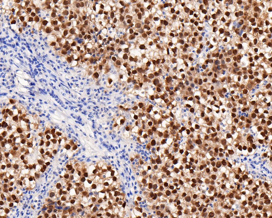
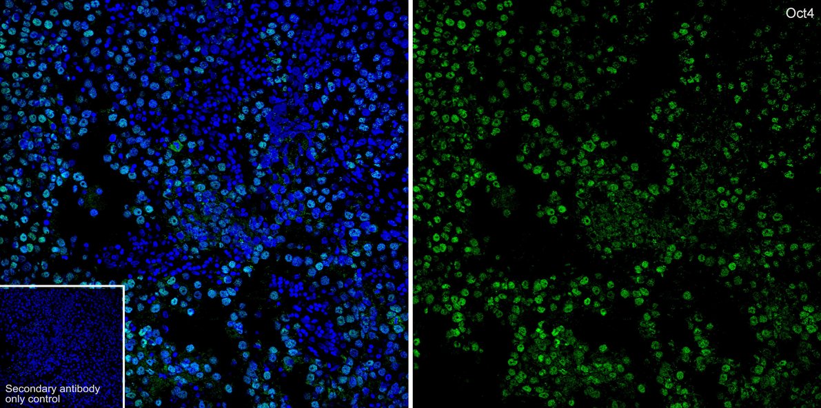
Immunohistochemical analysis of paraffin-embedded human seminoma tissue with Rabbit anti-Oct4 antibody (ET1612-20) at 1/4,000 dilution.
The section was pre-treated using heat mediated antigen retrieval with sodium citrate buffer (pH 6.0) for 2 minutes. The tissues were blocked in 1% BSA for 20 minutes at room temperature, washed with ddH2O and PBS, and then probed with the primary antibody (ET1612-20) at 1/4,000 dilution for 1 hour at room temperature. The detection was performed using an HRP conjugated compact polymer system. DAB was used as the chromogen. Tissues were counterstained with hematoxylin and mounted with DPX.

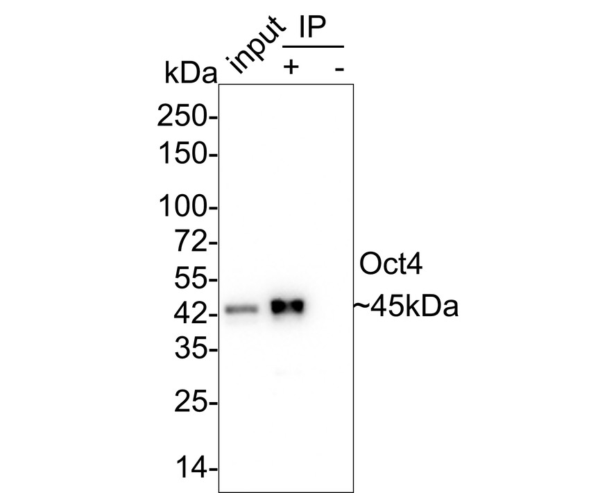
Oct4 was immunoprecipitated in 0.2mg NCCIT cell lysate with ET1612-20 at 2 µg/25 µl agarose. Western blot was performed from the immunoprecipitate using ET1612-20 at 1/2,000 dilution. Anti-Rabbit IgG for IP Nano-secondary antibody (NBI01H) at 1/5,000 dilution was used for 1 hour at room temperature.
Lane 1: NCCIT cell lysate (input)
Lane 2: ET1612-20 IP in NCCIT cell lysate
Lane 3: Rabbit IgG instead of ET1612-20 in NCCIT cell lysate
Blocking/Dilution buffer: 5% NFDM/TBST
Exposure time: 1 minute 40 seconds
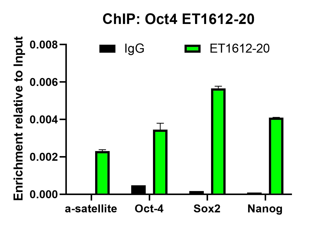
Chromatin immunoprecipitations were performed with cross-linked chromatin from NCCIT cells with Oct4 (ET1612-20) or Normal Rabbit IgG according to the ChIP protocol. The enriched DNA was quantified by real-time PCR using indicated primers. The amount of immunoprecipitated DNA in each sample is represented as signal relative to the total amount of input chromatin, which is equivalent to one.
Diminished GALNS activity in induced pluripotent stem cells of mucopolysaccharidosis IVA caused by compound p. S162Y and p. C165F mutation
Author: Jiang Xiaoling,et al
PMID: 39186005
期刊: Quarterly Journal Of Management
应用: IF
反应种属: Human
发表时间: 2024 Aug


Copyright © 广州杰特伟生物科技有限公司 All Rights Reserved. 备案号:粤ICP备19077843号