


Cyclin D1 Recombinant Rabbit Monoclonal Antibody [SA38-08]
Recombinant Rabbit monoclonal Antibody
Synthetic peptide within C-terminal human Cyclin D1.
Human, Mouse, Rat
WB, IF-Cell, IF-Tissue, IHC-P, IP, FC
Predicted band size: 34 kDa
MCF7 cell lysate, K-562 cell lysate, A431 cell lysate, Neuro-2a cell lysate, NIH/3T3 cell lysate, C6 cell lysate, SH-SY5Y cell lysate, Neuro-2a, MCF7, human tonsil tissue, human colon carcinoma tissue, human liver carcinoma tissue, human small intestine tissue.
unconjugated
SA38-08
Liquid
1ug/ul
Store at +4℃ after thawing. Aliquot store at -20℃ or -80℃. Avoid repeated freeze / thaw cycles.
1*TBS (pH7.4), 0.05% BSA, 40% Glycerol. Preservative: 0.05% Sodium Azide.
IgG
Protein A affinity purified.
WB
1:5,000
IF-Cell
1:2,000
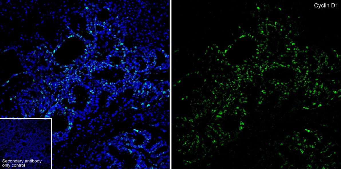
IF-Tissue
1:200-1:500
IHC-P
1:200-1:1,000
IP
Use at an assay dependent concentration.
FC
1:5,000
| Human | 查看 23 篇文献如下 |
| Mouse | 查看 11 篇文献如下 |
| Rat | 查看 1 篇文献如下 |
The protein encoded by this gene belongs to the highly conserved cyclin family, whose members are characterized by a dramatic periodicity in protein abundance throughout the cell cycle. Cyclins function as regulators of CDKs (Cyclin-dependent kinase). Different cyclins exhibit distinct expression and degradation patterns which contribute to the temporal coordination of each mitotic event. This cyclin forms a complex with and functions as a regulatory subunit of CDK4 or CDK6, whose activity is required for cell cycle G1/S transition. This protein has been shown to interact with tumor suppressor protein Rb and the expression of this gene is regulated positively by Rb. Mutations, amplification and overexpression of this gene, which alters cell cycle progression, are observed frequently in a variety of tumors and may contribute to tumorigenesis. Micrograph of cyclin D1 staining in a mantle cell lymphoma. Immunohistochemical staining of cyclin D1 antibodies is used to diagnose mantle cell lymphoma. Cyclin D1 has been found to be overexpressed in breast carcinoma. Its potential use as a biomarker was suggested.
1. Totta, P. et al. 2015. Clathrin heavy chain interacts with estrogen receptor α and modulates 17β-estradiol signaling. Molecular endocrinology (Baltimore, Md.). : me20141385.
2. Luo, Y. et al. 2015. Lycorine induces programmed necrosis in the multiple myeloma cell line ARH-77. Tumour biology : the journal of the International Society for Oncodevelopmental Biology and Medicine. 36: 2937-45.
Belongs to the cyclin family. Cyclin D subfamily.
Phosphorylation at Thr-286 by MAP kinases is required for ubiquitination and degradation following DNA damage. It probably plays an essential role for recognition by the FBXO31 component of SCF (SKP1-cullin-F-box) protein ligase complex.; Ubiquitinated, primarily as 'Lys-48'-linked polyubiquitination. Ubiquitinated by a SCF (SKP1-CUL1-F-box protein) ubiquitin-protein ligase complex containing FBXO4 and CRYAB. Following DNA damage it is ubiquitinated by some SCF (SKP1-cullin-F-box) protein ligase complex containing FBXO31. SCF-type ubiquitination is dependent on Thr-286 phosphorylation (By similarity). Ubiquitinated also by UHRF2 apparently in a phosphorylation-independent manner. Ubiquitination leads to its degradation and G1 arrest. Deubiquitinated by USP2; leading to its stabilization.
Cytoplasm, Nucleus, Membrane, Mitochondrion
AI327039 antibody
B cell CLL/lymphoma 1 antibody
B cell leukemia 1 antibody
B cell lymphoma 1 protein antibody
B-cell lymphoma 1 protein antibody
BCL 1 antibody
BCL-1 antibody
BCL-1 oncogene antibody
BCL1 antibody
BCL1 oncogene antibody
展开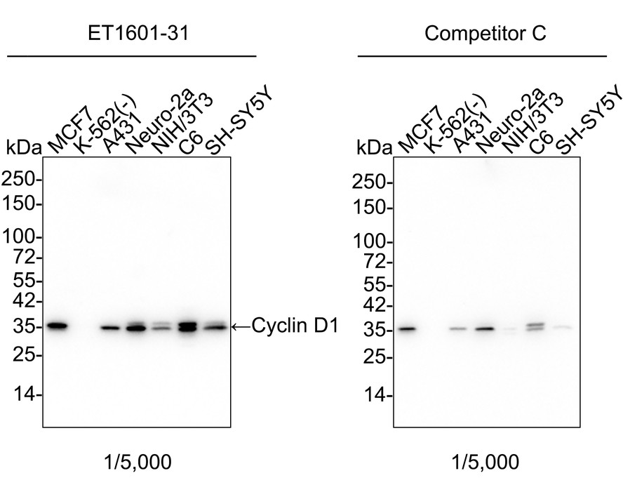
☑ Relative expression (RE)
Western blot analysis of Cyclin D1 on different lysates with Rabbit anti-Cyclin D1 antibody (ET1601-31) at 1/5,000 dilution and competitor's antibody at 1/5,000 dilution.
Lane 1: MCF7 cell lysate
Lane 2: K-562 cell lysate (negative)
Lane 3: A431 cell lysate
Lane 4: Neuro-2a cell lysate
Lane 5: NIH/3T3 cell lysate
Lane 6: C6 cell lysate
Lane 7: SH-SY5Y cell lysate
Lysates/proteins at 20 µg/Lane.
Predicted band size: 34 kDa
Observed band size: 35 kDa
Exposure time: 20 seconds; ECL: K1802;
4-20% SDS-PAGE gel.
Proteins were transferred to a PVDF membrane and blocked with 5% NFDM/TBST for 1 hour at room temperature. The primary antibody (ET1601-31) at 1/5,000 dilution and competitor's antibody at 1/5,000 dilution were used in 5% NFDM/TBST at 4℃ overnight. Goat Anti-Rabbit IgG - HRP Secondary Antibody (HA1001) at 1/50,000 dilution was used for 1 hour at room temperature.
☑ Knockdown (KD)
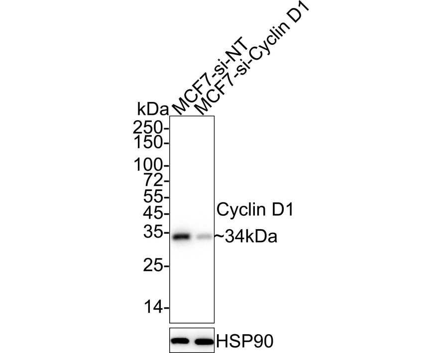
Western blot analysis of Cyclin D1 on different lysates with Rabbit anti-Cyclin D1 antibody (ET1601-31) at 1/5,000 dilution.
Lane 1: MCF7-si NT cell lysate
Lane 2: MCF7-si Cyclin D1 cell lysate
Lysates/proteins at 10 µg/Lane.
Predicted band size: 34 kDa
Observed band size: 34 kDa
Exposure time: 17 seconds; ECL: K1801;
4-20% SDS-PAGE gel.
Proteins were transferred to a PVDF membrane and blocked with 5% NFDM/TBST for 1 hour at room temperature. The primary antibody (ET1601-31) at 1/5,000 dilution was used in 5% NFDM/TBST at 4℃ overnight. Goat Anti-Rabbit IgG - HRP Secondary Antibody (HA1001) at 1/50,000 dilution was used for 1 hour at room temperature.

Cyclin D1 was immunoprecipitated from 0.5 mg Hela whole cell lysates with ET1601-31 at 2 μg/mL. Western blot was performed from the immunoprecipitate using ET1601-31 at 1/500 dilution for 45 minutes at room temperature. Goat anti-Rabbit IgG-HRP Secondary Antibody (HA1001) was used at 1:300,000 dilution for 30 minutes at room temperature.
Lane 1: Hela whole cell lysates at 10 μg;
Lane 2: Cyclin D1 (ET1601-31) IP in Hela whole cell lysates;
Lane 3: Rabbit IgG instead of Cyclin D1 (ET1601-31) in Hela whole cell lysates.
Predicted band size: 34 kDa
Observed band size: 34 kDa
Exposure time: 5 minutes;
12% SDS-PAGE gel.
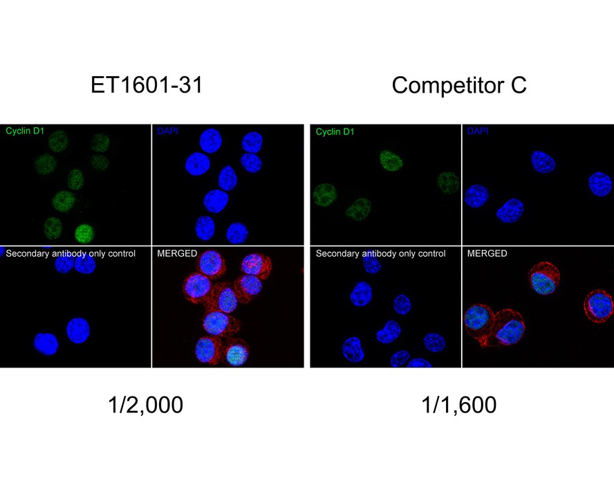
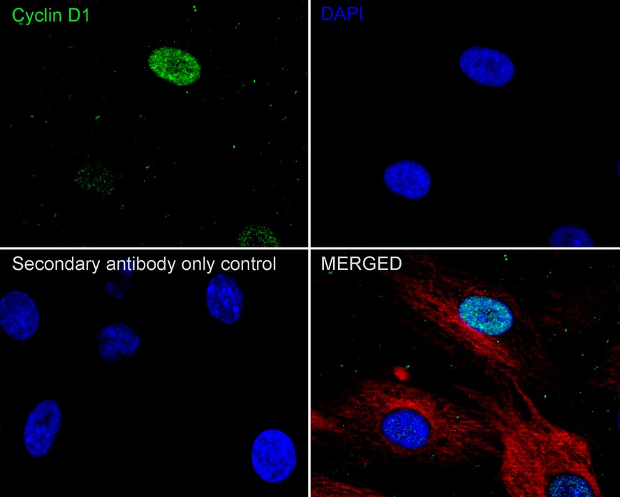
Immunocytochemistry analysis of Neuro-2a cells labeling Cyclin D1 with Rabbit anti-Cyclin D1 antibody (ET1601-31) at 1/2,000 dilution and competitor's antibody at 1/1,600 dilution.
Cells were fixed in 4% paraformaldehyde for 20 minutes at room temperature, permeabilized with 0.1% Triton X-100 in PBS for 5 minutes at room temperature, then blocked with 1% BSA in 10% negative goat serum for 1 hour at room temperature. Cells were then incubated with Rabbit anti-Cyclin D1 antibody (ET1601-31) at 1/2,000 dilution and competitor's antibody at 1/1,600 dilution in 1% BSA in PBST overnight at 4 ℃. Goat Anti-Rabbit IgG H&L (iFluor™ 488, HA1121) was used as the secondary antibody at 1/1,000 dilution. PBS instead of the primary antibody was used as the secondary antibody only control. Nuclear DNA was labelled in blue with DAPI.
Beta tubulin (M1305-2, red) was stained at 1/100 dilution overnight at +4℃. Goat Anti-Mouse IgG H&L (iFluor™ 594, HA1126) was used as the secondary antibody at 1/1,000 dilution.
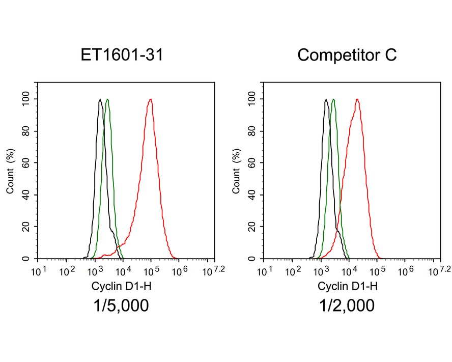
Flow cytometric analysis of MCF7 cells labeling Cyclin D1.
Cells were fixed and permeabilized. Then stained with the primary antibody (ET1601-31, red) at 1/5,000 dilution and competitor's antibody (red) at 1/2,000 dilution, compared with Rabbit IgG Isotype Control (green). After incubation of the primary antibody at +4℃ for an hour, the cells were stained with a iFluor™ 488 conjugate-Goat anti-Rabbit IgG Secondary antibody (HA1121) at 1/1,000 dilution for 30 minutes at +4℃. Unlabelled sample was used as a control (cells without incubation with primary antibody; black).
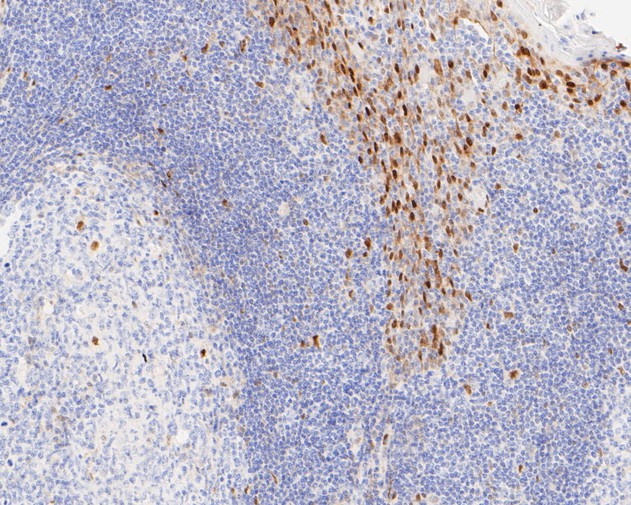
Immunohistochemical analysis of paraffin-embedded human tonsil tissue using anti-Cyclin D1 antibody. The section was pre-treated using heat mediated antigen retrieval with sodium citrate buffer (pH 6.0) for 2 minutes. The tissues were blocked in 5% BSA for 30 minutes at room temperature, washed with ddH2O and PBS, and then probed with the primary antibody (ET1601-31, 1/200) for 30 minutes at room temperature. The detection was performed using an HRP conjugated compact polymer system. DAB was used as the chromogen. Tissues were counterstained with hematoxylin and mounted with DPX.
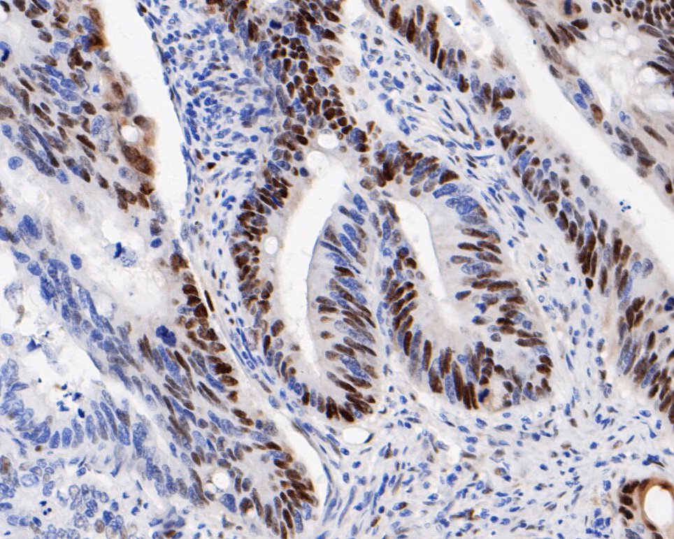
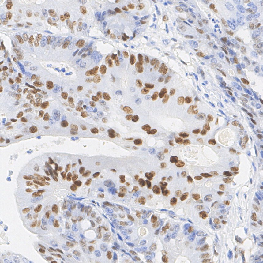
Immunohistochemical analysis of paraffin-embedded human colon carcinoma tissue using anti-Cyclin D1 antibody. The section was pre-treated using heat mediated antigen retrieval with sodium citrate buffer (pH 6.0) for 2 minutes. The tissues were blocked in 5% BSA for 30 minutes at room temperature, washed with ddH2O and PBS, and then probed with the primary antibody (ET1601-31, 1/200) for 30 minutes at room temperature. The detection was performed using an HRP conjugated compact polymer system. DAB was used as the chromogen. Tissues were counterstained with hematoxylin and mounted with DPX.
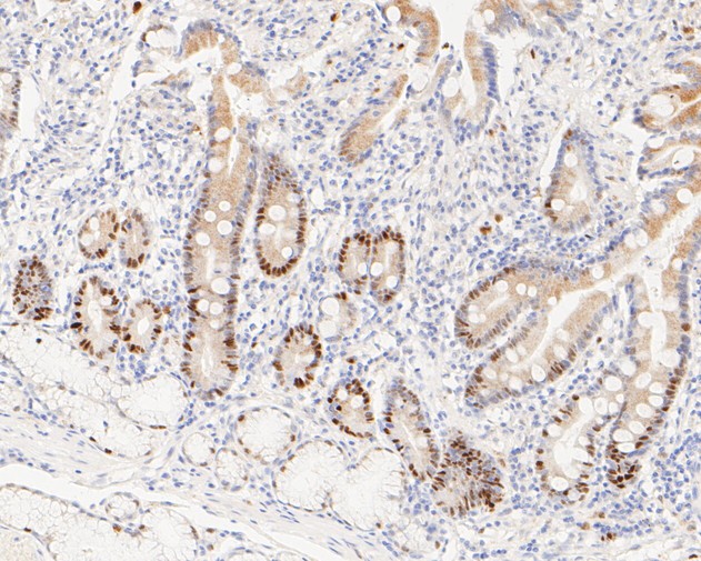
Immunohistochemical analysis of paraffin-embedded human small intestine tissue using anti-Cyclin D1 antibody. The section was pre-treated using heat mediated antigen retrieval with sodium citrate buffer (pH 6.0) for 2 minutes. The tissues were blocked in 5% BSA for 30 minutes at room temperature, washed with ddH2O and PBS, and then probed with the primary antibody (ET1601-31, 1/200) for 30 minutes at room temperature. The detection was performed using an HRP conjugated compact polymer system. DAB was used as the chromogen. Tissues were counterstained with hematoxylin and mounted with DPX.

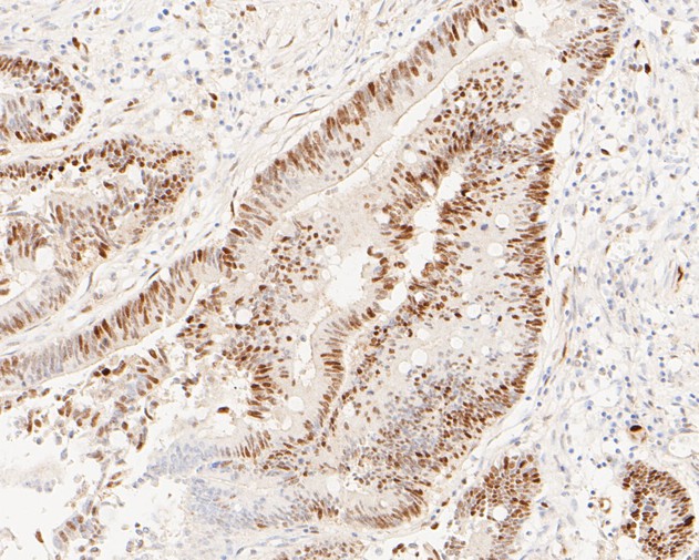
Immunohistochemical analysis of paraffin-embedded human colon carcinoma tissue using anti-Cyclin D1 antibody. The section was pre-treated using heat mediated antigen retrieval with sodium citrate buffer (pH 6.0) for 2 minutes. The tissues were blocked in 5% BSA for 30 minutes at room temperature, washed with ddH2O and PBS, and then probed with the primary antibody (ET1601-31, 1/200) for 30 minutes at room temperature. The detection was performed using an HRP conjugated compact polymer system. DAB was used as the chromogen. Tissues were counterstained with hematoxylin and mounted with DPX.
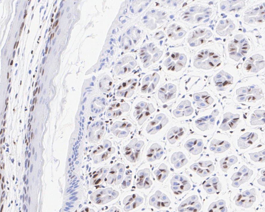
Immunohistochemical analysis of paraffin-embedded mouse stomach tissue with Rabbit anti-Cyclin D1 antibody (ET1601-31) at 1/1,000 dilution.
The section was pre-treated using heat mediated antigen retrieval with sodium citrate buffer (pH 6.0) for 2 minutes. The tissues were blocked in 1% BSA for 20 minutes at room temperature, washed with ddH2O and PBS, and then probed with the primary antibody (ET1601-31) at 1/1,000 dilution for 1 hour at room temperature. The detection was performed using an HRP conjugated compact polymer system. DAB was used as the chromogen. Tissues were counterstained with hematoxylin and mounted with DPX.
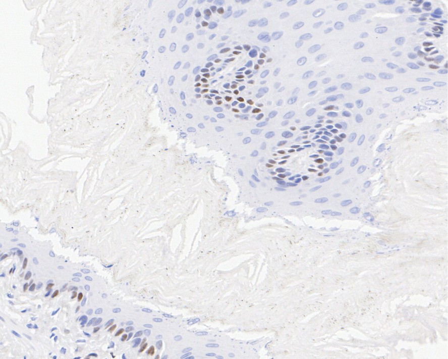
Immunohistochemical analysis of paraffin-embedded rat esophagus tissue with Rabbit anti-Cyclin D1 antibody (ET1601-31) at 1/1,000 dilution.
The section was pre-treated using heat mediated antigen retrieval with sodium citrate buffer (pH 6.0) for 2 minutes. The tissues were blocked in 1% BSA for 20 minutes at room temperature, washed with ddH2O and PBS, and then probed with the primary antibody (ET1601-31) at 1/1,000 dilution for 1 hour at room temperature. The detection was performed using an HRP conjugated compact polymer system. DAB was used as the chromogen. Tissues were counterstained with hematoxylin and mounted with DPX.
☑ Relative expression (RE)
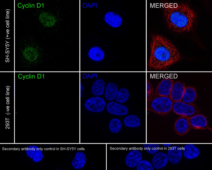
Immunocytochemistry analysis of SH-SY5Y (positive) and 293T (negative) labeling Cyclin D1 with Rabbit anti-Cyclin D1 antibody (ET1601-31) at 1/2,000 dilution.
Cells were fixed in 4% paraformaldehyde for 20 minutes at room temperature, permeabilized with 0.1% Triton X-100 in PBS for 5 minutes at room temperature, then blocked with 1% BSA in 10% negative goat serum for 1 hour at room temperature. Cells were then incubated with Rabbit anti-Cyclin D1 antibody (ET1601-31) at 1/2,000 dilution in 1% BSA in PBST overnight at 4 ℃. Goat Anti-Rabbit IgG H&L (iFluor™ 488, HA1121) was used as the secondary antibody at 1/1,000 dilution. PBS instead of the primary antibody was used as the secondary antibody only control. Nuclear DNA was labelled in blue with DAPI.
Beta tubulin (M1305-2, red) was stained at 1/100 dilution overnight at +4℃. Goat Anti-Mouse IgG H&L (iFluor™ 594, HA1126) was used as the secondary antibody at 1/1,000 dilution.

Immunocytochemistry analysis of C6 cells labeling Cyclin D1 with Rabbit anti-Cyclin D1 antibody (ET1601-31) at 1/2,000 dilution.
Cells were fixed in 4% paraformaldehyde for 20 minutes at room temperature, permeabilized with 0.1% Triton X-100 in PBS for 5 minutes at room temperature, then blocked with 1% BSA in 10% negative goat serum for 1 hour at room temperature. Cells were then incubated with Rabbit anti-Cyclin D1 antibody (ET1601-31) at 1/2,000 dilution in 1% BSA in PBST overnight at 4 ℃. Goat Anti-Rabbit IgG H&L (iFluor™ 488, HA1121) was used as the secondary antibody at 1/1,000 dilution. PBS instead of the primary antibody was used as the secondary antibody only control. Nuclear DNA was labelled in blue with DAPI.
Beta tubulin (M1305-2, red) was stained at 1/100 dilution overnight at +4℃. Goat Anti-Mouse IgG H&L (iFluor™ 594, HA1126) was used as the secondary antibody at 1/1,000 dilution.
Cyclin D1 was immunoprecipitated from 0.2 mg MCF7 cell lysate with ET1601-31 at 2 µg/25 µl agarose. Western blot was performed from the immunoprecipitate using ET1601-31 at 1/1,000 dilution. Anti-Rabbit IgG for IP Nano-secondary antibody (NBI01H) at 1/5,000 dilution was used for 1 hour at room temperature.
Lane 1: MCF7 cell lysate (input)
Lane 2: ET1601-31 IP in MCF7 cell lysate
Lane 3: Rabbit IgG instead of ET1601-31 in MCF7 cell lysate
Blocking/Dilution buffer: 5% NFDM/TBST
Exposure time: 5 seconds; ECL: K1802
Targeting NEDD8 in pediatric acute myeloid leukemia: an integrated bioinformatics and experimental approach
Author: Jian Sun, Cui Liu, Guangli Yang, Qian Li, Yang An, Yin Zhu, Pingping Zhang, Yaning Guan, Chang Peng, Zuochen Du, Pei Huang, Yan Chen
PMID: 40103351
期刊: Hematology
应用: WB
反应种属: Human
发表时间: 2025 Mar


Copyright © 广州杰特伟生物科技有限公司 All Rights Reserved. 备案号:粤ICP备19077843号