


PI 3 Kinase p85 alpha Recombinant Mouse Monoclonal Antibody [A3-D0-R]
Recombinant Mouse Monoclonal Antibody
Recombinant protein within Human PI3-kinase p85 subunit alpha aa 19-219 / 724.
Human
WB, IF-Cell
Predicted band size: 84 kDa
SW480 cell lysate, A431 cell lysate, 293T cell lysate, Jurkat cell lysate, Raji cell lysate, MCF7 cell lysate, A549 cell lysate, U-937.
unconjugated
A3-D0-R
Liquid
1ug/ul
Store at +4℃ after thawing. Aliquot store at -20℃. Avoid repeated freeze / thaw cycles.
PBS (pH7.4), 0.1% BSA, 40% Glycerol. Preservative: 0.05% Sodium Azide.
IgG1
Protein A affinity purified.
WB
1:2,000-1:5,000
IF-Cell
1:100
| Rat | 查看 1 篇文献如下 |
| Human | 查看 1 篇文献如下 |
| Human | 查看 1 篇文献如下 |
Phosphatidylinositol 3-kinase (PI 3-kinase) is composed of (p85) and (p110) subunits. p85 lacks PI 3-kinase activity and acts as an adapter, coupling p110 to activated protein tyrosine kinase. Two forms of p85 have been described (p85α and p85β), each possessing one SH3 and two SH2 domains. Various p110 isoforms have been identified. p110α and p110β interact with p85α, and p110α has also been shown to interact with p85β in vitro. p110δ expression is restricted to white blood cells. It has been shown to bind p85α and γ, but it apparently does not phosphorylate these subunits. p110δ seems to have the capacity to autophosphorylate. p110γ does not interact with the p85 subunits. It has been shown to be activated by α and βγ heterotrimeric G proteins.
1. Hu J et al. Filamin B regulates chondrocyte proliferation and differentiation through Cdk1 signaling. PLoS One 9:e89352 (2014).
2. Schmidt JW et al. Stat5 regulates the phosphatidylinositol 3-kinase/Akt1 pathway during mammary gland development and tumorigenesis. Mol Cell Biol 34:1363-77 (2014).
Cytosol, Membrane, Nucleus.
GRB1 antibody
p85 alpha antibody
p85 antibody
P85A_HUMAN antibody
Phosphatidylinositol 3 kinase associated p 85 alpha antibody
Phosphatidylinositol 3 kinase regulatory 1 antibody
Phosphatidylinositol 3 kinase, regulatory subunit, polypeptide 1 (p85 alpha) antibody
Phosphatidylinositol 3-kinase 85 kDa regulatory subunit alpha antibody
Phosphatidylinositol 3-kinase regulatory subunit alpha antibody
Phosphoinositide 3 kinase, regulatory subunit 1 (alpha) antibody
展开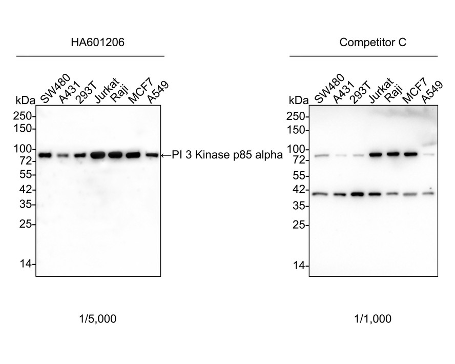
Western blot analysis of PI 3 Kinase p85 alpha on different lysates with Mouse anti-PI 3 Kinase p85 alpha antibody (HA601206) at 1/5,000 dilution and competitor's antibody at 1/1,000 dilution.
Lane 1: SW480 cell lysate
Lane 2: A431 cell lysate
Lane 3: 293T cell lysate
Lane 4: Jurkat cell lysate
Lane 5: Raji cell lysate
Lane 6: MCF7 cell lysate
Lane 7: A549 cell lysate
Lysates/proteins at 15 µg/Lane.
Predicted band size: 84 kDa
Observed band size: 84 kDa
Exposure time: 1 minute 40 seconds; ECL: K1802;
4-20% SDS-PAGE gel.
Proteins were transferred to a PVDF membrane and blocked with 5% NFDM/TBST for 1 hour at room temperature. The primary antibody (HA601206) at 1/5,000 dilution and competitor's antibody at 1/1,000 dilution were used in 5% NFDM/TBST at 4℃ overnight. Goat Anti-Mouse IgG - HRP Secondary Antibody (HA1006) at 1/50,000 dilution was used for 1 hour at room temperature.
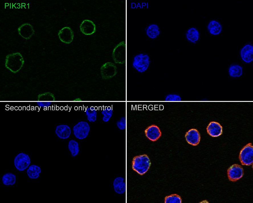
Immunocytochemistry analysis of U-937 cells labeling PI 3 Kinase p85 alpha with Mouse anti-PI 3 Kinase p85 alpha antibody (HA601206) at 1/100 dilution.
Cells were fixed in 4% paraformaldehyde for 20 minutes at room temperature, permeabilized with 0.1% Triton X-100 in PBS for 5 minutes at room temperature, then blocked with 1% BSA in 10% negative goat serum for 1 hour at room temperature. Cells were then incubated with Mouse anti-PI 3 Kinase p85 alpha antibody (HA601206) at 1/100 dilution in 1% BSA in PBST overnight at 4 ℃. Goat Anti-Mouse IgG H&L (iFluor™ 488, HA1125) was used as the secondary antibody at 1/1,000 dilution. PBS instead of the primary antibody was used as the secondary antibody only control. Nuclear DNA was labelled in blue with DAPI.
beta Tubulin (ET1602-4, red) was stained at 1/100 dilution overnight at +4℃. Goat Anti-Rabbit IgG H&L (iFluor™ 594, HA1122) were used as the secondary antibody at 1/1,000 dilution.

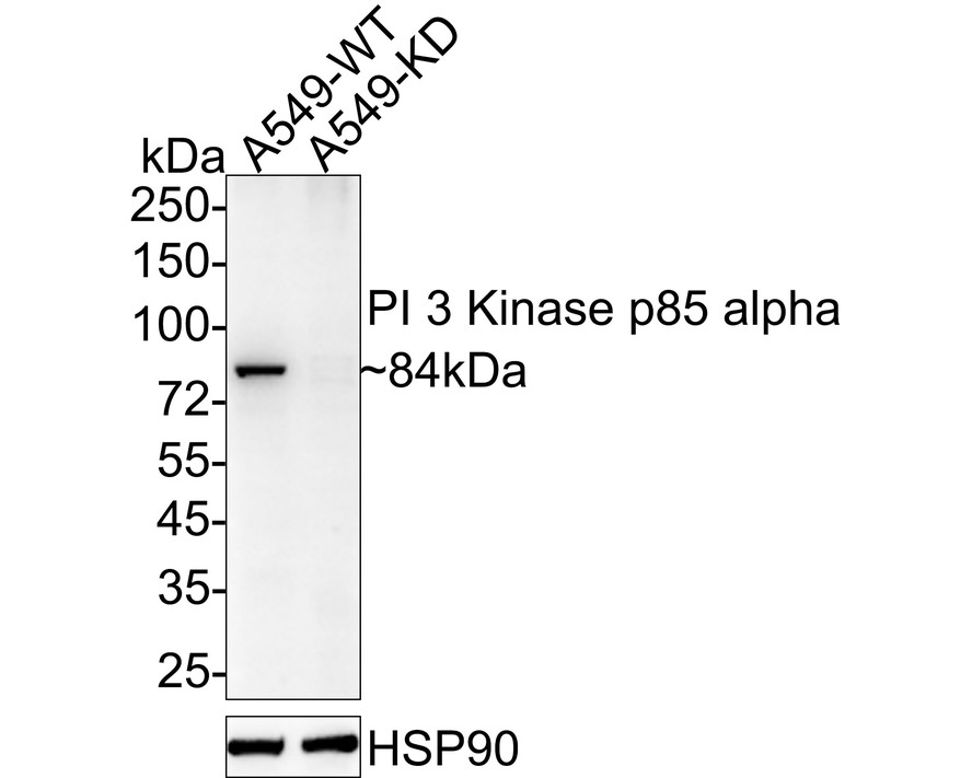
☑ Knockdown (KD)
Western blot analysis of PI 3 Kinase p85 alpha on different lysates with Mouse anti-PI 3 Kinase p85 alpha antibody (HA601206) at 1/5,000 dilution.
Lane 1: A549-si NT cell lysate
Lane 2: A549-si PI 3 Kinase p85 alpha cell lysate
Lysates/proteins at 20 µg/Lane.
Predicted band size: 84 kDa
Observed band size: 84 kDa
Exposure time: 2 minutes; ECL: K1802;
4-20% SDS-PAGE gel.
Proteins were transferred to a PVDF membrane and blocked with 5% NFDM/TBST for 1 hour at room temperature. The primary antibody (HA601206) at 1/5,000 dilution was used in 5% NFDM/TBST at 4℃ overnight. Goat Anti-Mouse IgG - HRP Secondary Antibody (HA1006) at 1/50,000 dilution was used for 1 hour at room temperature.
Icariin alleviates osteoarthritis through PI3K/Akt/mTOR/ULK1 signaling pathway
Author:
PMID: 36253872
期刊: European Journal Of Medical Research
应用: WB
反应种属: Human
发表时间: 2022 Oct


Copyright © 广州杰特伟生物科技有限公司 All Rights Reserved. 备案号:粤ICP备19077843号