


Phospho-Met (Y1349) Recombinant Rabbit Monoclonal Antibody [JE51-68]
Recombinant Rabbit monoclonal Antibody
Synthetic phospho-peptide corresponding to residues surrounding Tyr1,349 of human Met.
Human, Rat
WB, IHC-P
Predicted band size: 156 kDa
HeLa treated with 100ng/mL Calyculin A for 30 minutes cell lysate, PC-12, human colon carcinoma tissue, human small intestine tissue, human rectum tissue.
unconjugated
JE51-68
Liquid
1ug/ul
Store at +4℃ after thawing. Aliquot store at -20℃. Avoid repeated freeze / thaw cycles.
1*TBS (pH7.4), 0.05% BSA, 40% Glycerol. Preservative: 0.05% Sodium Azide.
IgG
Protein A affinity purified.
WB
1:1,000-1:2,000
IHC-P
1:50-1:200
Receptor tyrosine kinase that transduces signals from the extracellular matrix into the cytoplasm by binding to hepatocyte growth factor/HGF ligand. Regulates many physiological processes including proliferation, scattering, morphogenesis and survival. Ligand binding at the cell surface induces autophosphorylation of MET on its intracellular domain that provides docking sites for downstream signaling molecules. Following activation by ligand, interacts with the PI3-kinase subunit PIK3R1, PLCG1, SRC, GRB2, STAT3 or the adapter GAB1. Recruitment of these downstream effectors by MET leads to the activation of several signaling cascades including the RAS-ERK, PI3 kinase-AKT, or PLCgamma-PKC. The RAS-ERK activation is associated with the morphogenetic effects while PI3K/AKT coordinates prosurvival effects. During embryonic development, MET signaling plays a role in gastrulation, development and migration of muscles and neuronal precursors, angiogenesis and kidney formation. In adults, participates in wound healing as well as organ regeneration and tissue remodeling. Promotes also differentiation and proliferation of hematopoietic cells. May regulate cortical bone osteogenesis (By similarity).
1. Niemann H.H. et. al. Structure of the human receptor tyrosine kinase Met in complex with the Listeria invasion protein InlB. Cell 130:235-246(2007).
2. Ferraris D.M. et. al. Ligand-mediated dimerization of the Met receptor tyrosine kinase by the bacterial invasion protein InlB. J. Mol. Biol. 395:522-532(2010).
Belongs to the protein kinase superfamily. Tyr protein kinase family.
Expressed in normal hepatocytes as well as in epithelial cells lining the stomach, the small and the large intestine. Found also in basal keratinocytes of esophagus and skin. High levels are found in liver, gastrointestinal tract, thyroid and kidney. Also present in the brain. Expressed in metaphyseal bone (at protein level).
Autophosphorylated in response to ligand binding on Tyr-1234 and Tyr-1235 in the kinase domain leading to further phosphorylation of Tyr-1349 and Tyr-1356 in the C-terminal multifunctional docking site. Dephosphorylated by PTPRJ at Tyr-1349 and Tyr-1365. Dephosphorylated by PTPN1 and PTPN2.; Ubiquitinated. Ubiquitination by CBL regulates MET endocytosis, resulting in decreasing plasma membrane receptor abundance, and in endosomal degradation and/or recycling of internalized receptors.; (Microbial infection) Tyrosine phosphorylation is stimulated by L.monocytogenes InlB. Tyrosine phosphorylation is maximal 10-20 minutes after treatment with InlB and disappears by 60 minutes. The phosphorylated residues were not identified.
Membrane, secreted.
AUTS9 antibody
c met antibody
D249 antibody
Hepatocyte growth factor receptor antibody
HGF antibody
HGF receptor antibody
HGF/SF receptor antibody
HGFR antibody
MET antibody
Met proto oncogene tyrosine kinase antibody
展开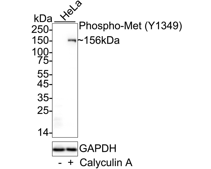
☑ Cell treatment (CT)
Western blot analysis of Phospho-Met (Y1349) on different lysates with Rabbit anti-Phospho-Met (Y1349) antibody (ET7110-14) at 1/1,000 dilution.
Lane 1: HeLa cell lysate
Lane 2: HeLa treated with 100ng/mL Calyculin A for 30 minutes cell lysate
Lysates/proteins at 20 µg/Lane.
Predicted band size: 156 kDa
Observed band size: 156 kDa
Exposure time: 1 minute; ECL: K1801;
4-20% SDS-PAGE gel.
Proteins were transferred to a PVDF membrane and blocked with 5% NFDM/TBST for 1 hour at room temperature. The primary antibody (ET7110-14) at 1/1,000 dilution was used in 5% NFDM/TBST at 4℃ overnight. Goat Anti-Rabbit IgG - HRP Secondary Antibody (HA1001) at 1/50,000 dilution was used for 1 hour at room temperature.

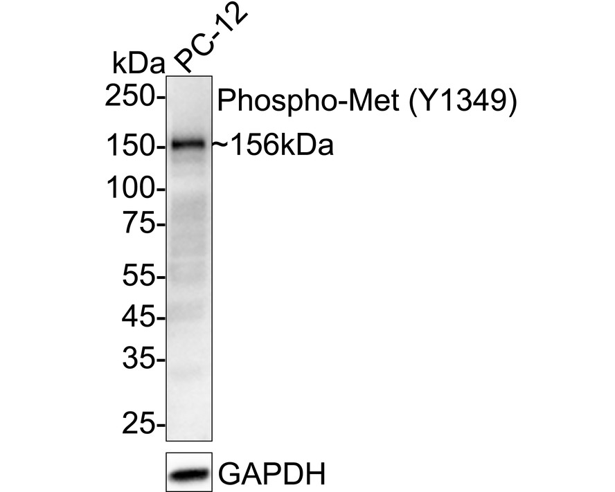
Western blot analysis of Phospho-Met (Y1349) on PC-12 cell lysates with Rabbit anti-Phospho-Met (Y1349) antibody (ET7110-14) at 1/2,000 dilution.
Lysates/proteins at 20 µg/Lane.
Predicted band size: 156 kDa
Observed band size: 156 kDa
Exposure time: 25 seconds; ECL: K1802;
4-20% SDS-PAGE gel.
Proteins were transferred to a PVDF membrane and blocked with 5% NFDM/TBST for 1 hour at room temperature. The primary antibody (ET7110-14) at 1/2,000 dilution was used in 5% NFDM/TBST at 4℃ overnight. Goat Anti-Rabbit IgG - HRP Secondary Antibody (HA1001) at 1/50,000 dilution was used for 1 hour at room temperature.
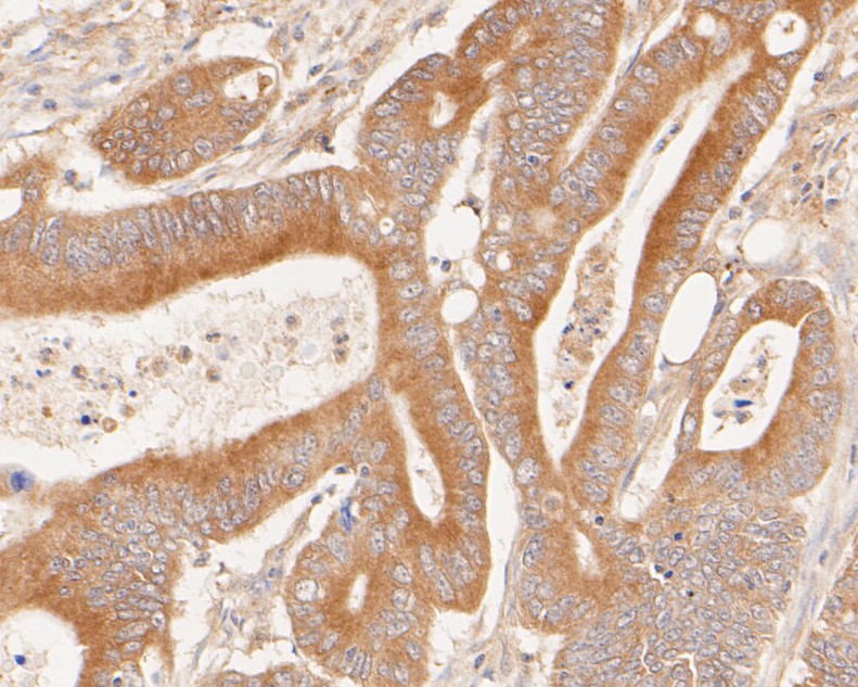
Immunohistochemical analysis of paraffin-embedded human colon carcinoma tissue using anti-Phospho-Met (Y1349) antibody. The section was pre-treated using heat mediated antigen retrieval with Tris-EDTA buffer (pH 8.0-8.4) for 20 minutes.The tissues were blocked in 5% BSA for 30 minutes at room temperature, washed with ddH2O and PBS, and then probed with the primary antibody (ET7110-14, 1/50) for 30 minutes at room temperature. The detection was performed using an HRP conjugated compact polymer system. DAB was used as the chromogen. Tissues were counterstained with hematoxylin and mounted with DPX.
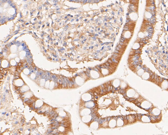
Immunohistochemical analysis of paraffin-embedded human small intestine tissue using anti-Phospho-Met (Y1349) antibody. The section was pre-treated using heat mediated antigen retrieval with Tris-EDTA buffer (pH 8.0-8.4) for 20 minutes.The tissues were blocked in 5% BSA for 30 minutes at room temperature, washed with ddH2O and PBS, and then probed with the primary antibody (ET7110-14, 1/50) for 30 minutes at room temperature. The detection was performed using an HRP conjugated compact polymer system. DAB was used as the chromogen. Tissues were counterstained with hematoxylin and mounted with DPX.
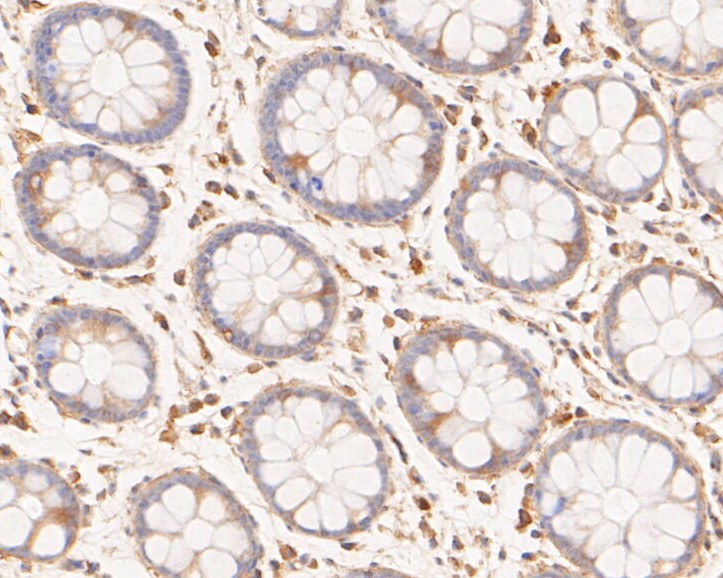
Immunohistochemical analysis of paraffin-embedded human rectum tissue using anti-Phospho-Met (Y1349) antibody. The section was pre-treated using heat mediated antigen retrieval with Tris-EDTA buffer (pH 8.0-8.4) for 20 minutes.The tissues were blocked in 5% BSA for 30 minutes at room temperature, washed with ddH2O and PBS, and then probed with the primary antibody (ET7110-14, 1/50) for 30 minutes at room temperature. The detection was performed using an HRP conjugated compact polymer system. DAB was used as the chromogen. Tissues were counterstained with hematoxylin and mounted with DPX.


Copyright © 广州杰特伟生物科技有限公司 All Rights Reserved. 备案号:粤ICP备19077843号