


ATG5 Recombinant Rabbit Monoclonal Antibody [SN73-07]
Recombinant Rabbit monoclonal Antibody
Synthetic peptide within Human ATG5 aa 1-50 / 275.
Human, Mouse, Rat, Monkey
WB, IF-Cell, IF-Tissue, IHC-P, IP, FC
Predicted band size: 32 kDa
NIH/3T3 cell lysate, C2C12 cell lysate, Neuro-2a cell lysate, PC-12 cell lysate, mouse brain tissue lysate, rat brain tissue lysate, HeLa cell lysate, A431 cell lysate, human brain tissue, NIH/3T3, PC-12.
unconjugated
SN73-07
Liquid
1ug/ul
Store at +4℃ after thawing. Aliquot store at -20℃ or -80℃. Avoid repeated freeze / thaw cycles.
1*TBS (pH7.4), 0.05% BSA, 40% Glycerol. Preservative: 0.05% Sodium Azide.
IgG
Protein A affinity purified.
WB
1:5,000-1:10,000
IF-Cell
1:100-1:250
IF-Tissue
1:50-1:200
IHC-P
1:2,000
IP
1-2μg/sample
FC
1:1,000
| Mouse | 查看 14 篇文献如下 |
| Human | 查看 14 篇文献如下 |
| Rat | 查看 2 篇文献如下 |
Involved in autophagic vesicle formation. Conjugation with ATG12, through a ubiquitin-like conjugating system involving ATG7 as an E1-like activating enzyme and ATG10 as an E2-like conjugating enzyme, is essential for its function. The ATG12-ATG5 conjugate acts as an E3-like enzyme which is required for lipidation of ATG8 family proteins and their association to the vesicle membranes. Involved in mitochondrial quality control after oxidative damage, and in subsequent cellular longevity. Plays a critical role in multiple aspects of lymphocyte development and is essential for both B and T lymphocyte survival and proliferation. Required for optimal processing and presentation of antigens for MHC II. Involved in the maintenance of axon morphology and membrane structures, as well as in normal adipocyte differentiation. Promotes primary ciliogenesis through removal of OFD1 from centriolar satellites and degradation of IFT20 via the autophagic pathway.
1. He G et al. Gadd45b prevents autophagy and apoptosis against rat cerebral neuron oxygen-glucose deprivation/reperfusion injury. Apoptosis 21:390-403 (2016).
2. Pla A et al. TLR4 mediates the impairment of ubiquitin-proteasome and autophagy-lysosome pathways induced by ethanol treatment in brain. Cell Death Dis 5:e1066 (2014).
Belongs to the ATG5 family.
Ubiquitous. The mRNA is present at similar levels in viable and apoptotic cells, whereas the protein is dramatically highly expressed in apoptotic cells.
Conjugated to ATG12; which is essential for autophagy, but is not required for association with isolation membrane.; Acetylated by EP300.
Cytoplasm, Membrane.
APG 5 antibody
APG 5L antibody
APG5 antibody
APG5 autophagy 5 like antibody
APG5 like antibody
APG5-like antibody
APG5L antibody
Apoptosis specific protein antibody
Apoptosis-specific protein antibody
ASP antibody
展开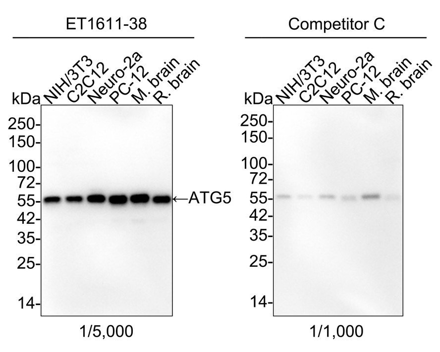
Western blot analysis of ATG5 on different lysates with Rabbit anti-ATG5 antibody (ET1611-38) at 1/5,000 dilution and competitor's antibody at 1/1,000 dilution.
Lane 1: NIH/3T3 cell lysate (15 µg/Lane)
Lane 2: C2C12 cell lysate (15 µg/Lane)
Lane 3: Neuro-2a cell lysate (15 µg/Lane)
Lane 4: PC-12 cell lysate (15 µg/Lane)
Lane 5: Mouse brain tissue lysate (15 µg/Lane)
Lane 6: Rat brain tissue lysate (15 µg/Lane)
Predicted band size: 32 kDa
Observed band size: 55 kDa
Exposure time: 35 seconds; ECL: K1802;
4-20% SDS-PAGE gel.
Proteins were transferred to a PVDF membrane and blocked with 5% NFDM/TBST for 1 hour at room temperature. The primary antibody (ET1611-38) at 1/5,000 dilution and competitor's antibody at 1/1,000 dilution were used in 5% NFDM/TBST at 4℃ overnight. Goat Anti-Rabbit IgG - HRP Secondary Antibody (HA1001) at 1/50,000 dilution was used for 1 hour at room temperature.
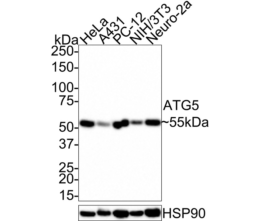
Western blot analysis of ATG5 on different lysates with Rabbit anti-ATG5 antibody (ET1611-38) at 1/5,000 dilution.
Lane 1: HeLa cell lysate (20 µg/Lane)
Lane 2: A431 cell lysate (20 µg/Lane)
Lane 3: PC-12 cell lysate (20 µg/Lane)
Lane 4: NIH/3T3 cell lysate (20 µg/Lane)
Lane 5: Neuro-2a cell lysate (20 µg/Lane)
Predicted band size: 32 kDa
Observed band size: 55 kDa
Exposure time: 1 minute; ECL: K1801;
4-20% SDS-PAGE gel.
Proteins were transferred to a PVDF membrane and blocked with 5% NFDM/TBST for 1 hour at room temperature. The primary antibody (ET1611-38) at 1/5,000 dilution was used in 5% NFDM/TBST at room temperature for 2 hours. Goat Anti-Rabbit IgG - HRP Secondary Antibody (HA1001) at 1/100,000 dilution was used for 1 hour at room temperature.
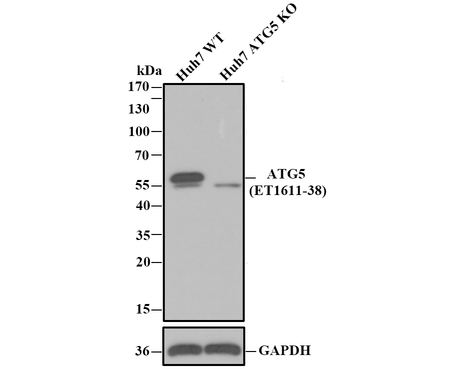
☑ Knockout (KO)
Western blot analysis of ATG5 with anti-ATG5 antibody [SN73-07] (ET1611-38) at 1/1,000 dilution.
Lane 1: Wild-type Huh7 whole cell lysate.
Lane 2: ATG5 knockout Huh7 whole cell lysate.
Proteins were transferred to a PVDF membrane and blocked with 5% NFDM in TBST for 1 hour at room temperature. The primary Anti-ATG5 antibody (ET1611-38, 1/1,000) and Anti-GAPDH antibody (ET1601-4, 1/10,000) were used in 5% BSA at room temperature for 2 hours. Goat Anti-Rabbit IgG H&L (HRP) Secondary Antibody (HA1001) at 1/200,000 dilution was used for 1 hour at room temperature.
Cell lysate was provided by Ubigene Biosciences (Ubigene Biosciences Co., Ltd., Guangzhou, China).
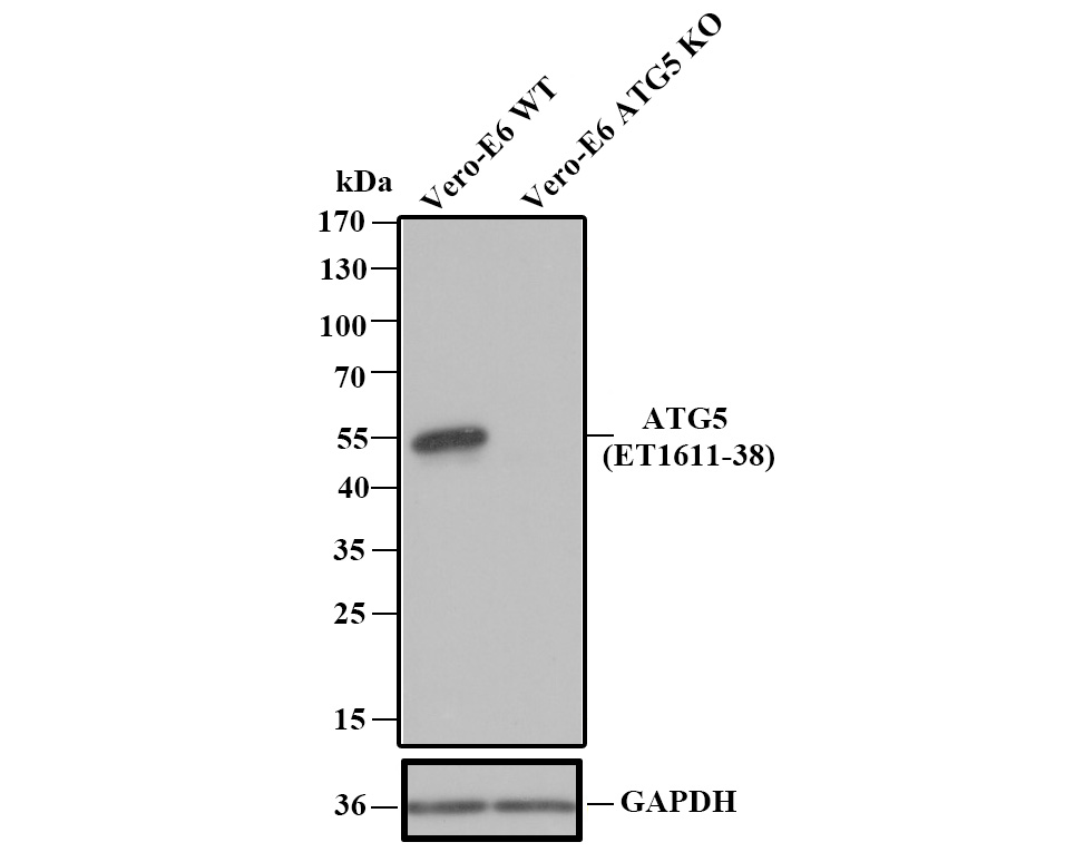
☑ Knockout (KO)
Western blot analysis of ATG5 with anti-ATG5 antibody [SN73-07] (ET1611-38) at 1/1,000 dilution.
Lane 1: Wild-type Vero-E6 whole cell lysate.
Lane 2: ATG5 knockout Vero-E6 whole cell lysate.
Proteins were transferred to a PVDF membrane and blocked with 5% NFDM in TBST for 1 hour at room temperature. The primary Anti-ATG5 antibody (ET1611-38, 1/1,000) and Anti-GAPDH antibody (ET1601-4, 1/10,000) were used in 5% BSA at room temperature for 2 hours. Goat Anti-Rabbit IgG H&L (HRP) Secondary Antibody (HA1001) at 1/200,000 dilution was used for 1 hour at room temperature.
Cell lysate was provided by Ubigene Biosciences (Ubigene Biosciences Co., Ltd., Guangzhou, China).
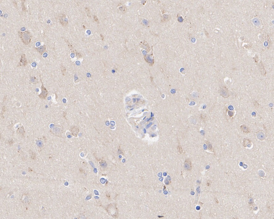
Immunohistochemical analysis of paraffin-embedded human brain tissue with Rabbit anti-ATG5 antibody (ET1611-38) at 1/2,000 dilution.
The section was pre-treated using heat mediated antigen retrieval with Tris-EDTA buffer (pH 9.0) for 20 minutes. The tissues were blocked in 1% BSA for 20 minutes at room temperature, washed with ddH2O and PBS, and then probed with the primary antibody (ET1611-38) at 1/2,000 dilution for 1 hour at room temperature. The detection was performed using an HRP conjugated compact polymer system. DAB was used as the chromogen. Tissues were counterstained with hematoxylin and mounted with DPX.
Immunocytochemistry analysis of NIH/3T3 cells labeling ATG5 with Rabbit anti-ATG5 antibody (ET1611-38) at 1/250 dilution.
Cells were fixed in 4% paraformaldehyde for 20 minutes at room temperature, permeabilized with 0.1% Triton X-100 in PBS for 5 minutes at room temperature, then blocked with 1% BSA in 10% negative goat serum for 1 hour at room temperature. Cells were then incubated with Rabbit anti-ATG5 antibody (ET1611-38) at 1/250 dilution in 1% BSA in PBST overnight at 4 ℃. Goat Anti-Rabbit IgG H&L (iFluor™ 488, HA1121) was used as the secondary antibody at 1/1,000 dilution. PBS instead of the primary antibody was used as the secondary antibody only control. Nuclear DNA was labelled in blue with DAPI. Beta tubulin (M1305-2, red) was stained at 1/100 dilution overnight at +4℃. Goat Anti-Mouse IgG H&L (iFluor™ 594, HA1126) was used as the secondary antibody at 1/1,000 dilution.

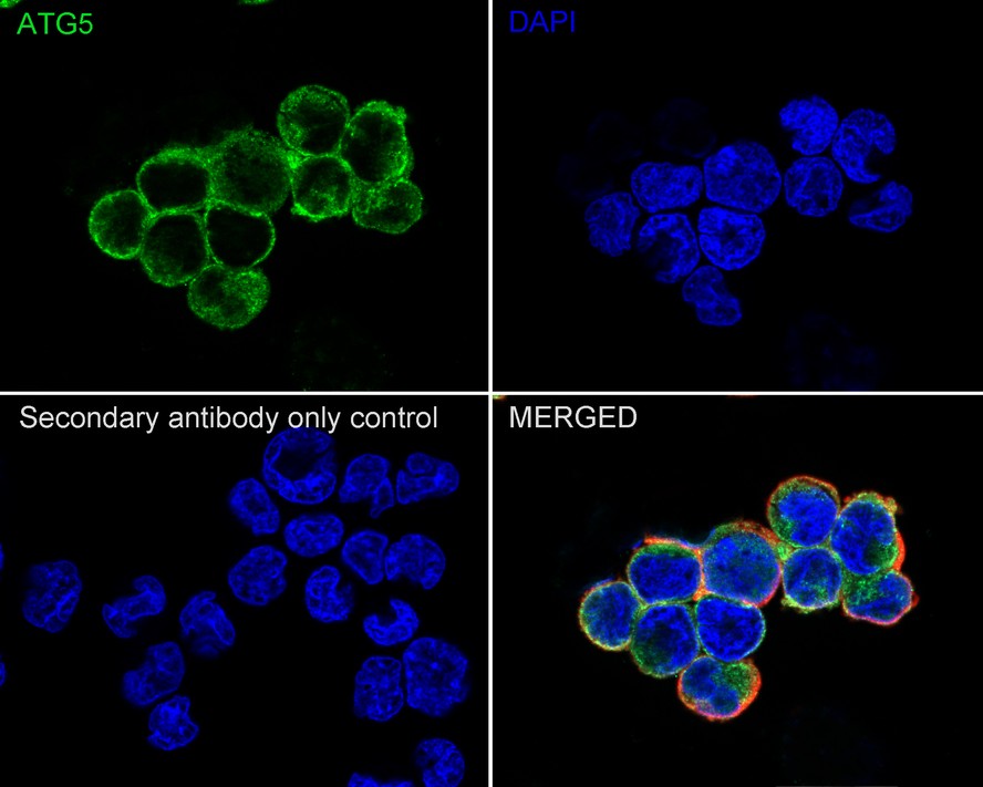
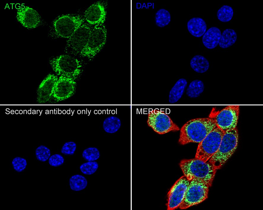
Immunocytochemistry analysis of PC-12 cells labeling ATG5 with Rabbit anti-ATG5 antibody (ET1611-38) at 1/100 dilution.
Cells were fixed in 4% paraformaldehyde for 20 minutes at room temperature, permeabilized with 0.1% Triton X-100 in PBS for 5 minutes at room temperature, then blocked with 1% BSA in 10% negative goat serum for 1 hour at room temperature. Cells were then incubated with Rabbit anti-ATG5 antibody (ET1611-38) at 1/100 dilution in 1% BSA in PBST overnight at 4 ℃. Goat Anti-Rabbit IgG H&L (iFluor™ 488, HA1121) was used as the secondary antibody at 1/1,000 dilution. PBS instead of the primary antibody was used as the secondary antibody only control. Nuclear DNA was labelled in blue with DAPI. Beta tubulin (M1305-2, red) was stained at 1/100 dilution overnight at +4℃. Goat Anti-Mouse IgG H&L (iFluor™ 594, HA1126) was used as the secondary antibody at 1/1,000 dilution.
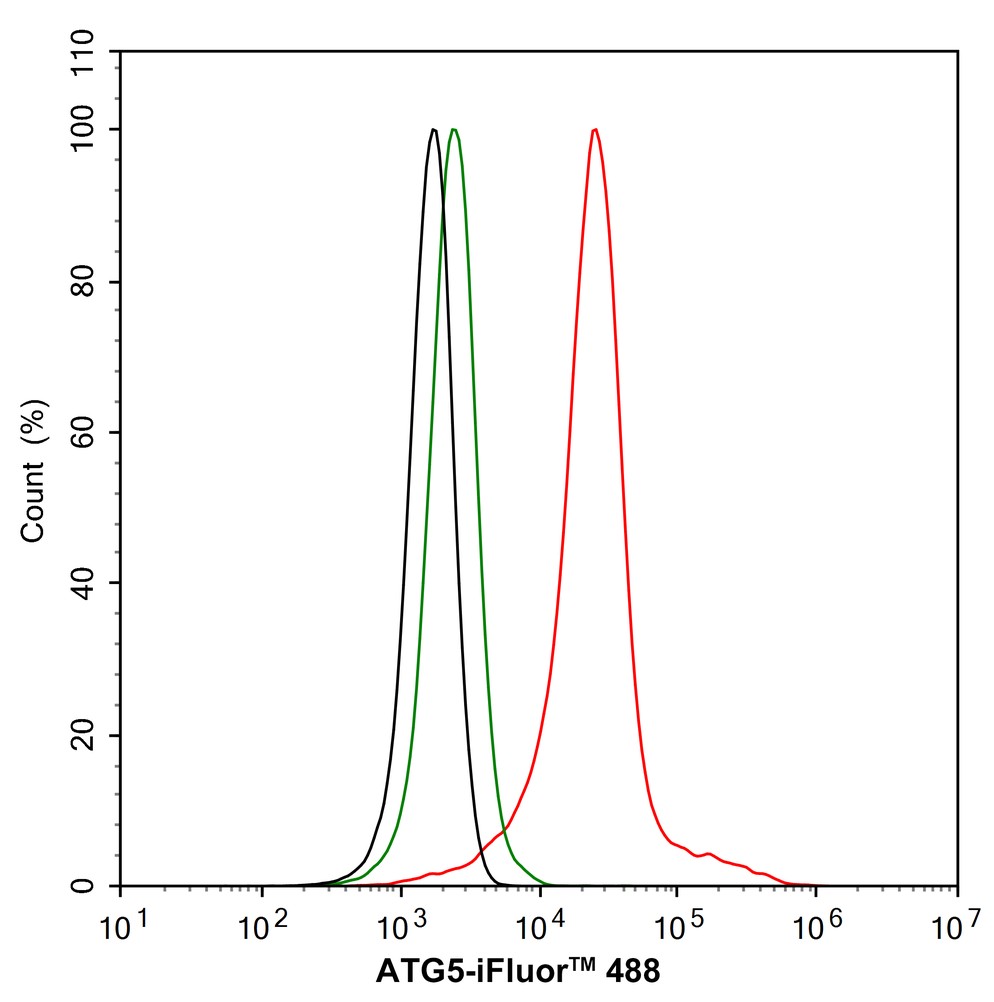
Flow cytometric analysis of NIH/3T3 cells labeling ATG5.
Cells were fixed and permeabilized. Then stained with the primary antibody (ET1611-38, 1/1,000) (red) compared with Rabbit IgG Isotype Control (green). After incubation of the primary antibody at +4℃ for an hour, the cells were stained with a iFluor™ 488 conjugate-Goat anti-Rabbit IgG Secondary antibody (HA1121) at 1/1,000 dilution for 30 minutes at +4℃. Unlabelled sample was used as a control (cells without incubation with primary antibody; black).
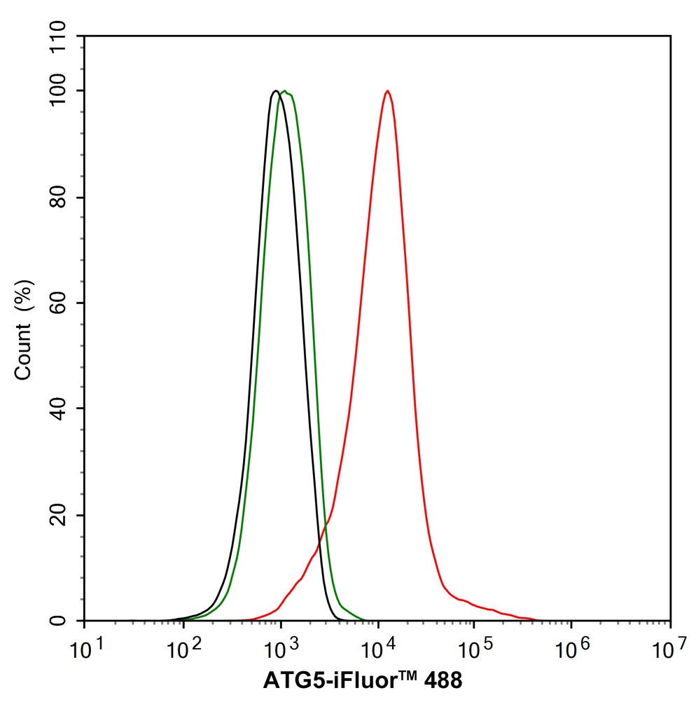
Flow cytometric analysis of PC-12 cells labeling ATG5.
Cells were fixed and permeabilized. Then stained with the primary antibody (ET1611-38, 1/1,000) (red) compared with Rabbit IgG Isotype Control (green). After incubation of the primary antibody at +4℃ for an hour, the cells were stained with a iFluor™ 488 conjugate-Goat anti-Rabbit IgG Secondary antibody (HA1121) at 1/1,000 dilution for 30 minutes at +4℃. Unlabelled sample was used as a control (cells without incubation with primary antibody; black).

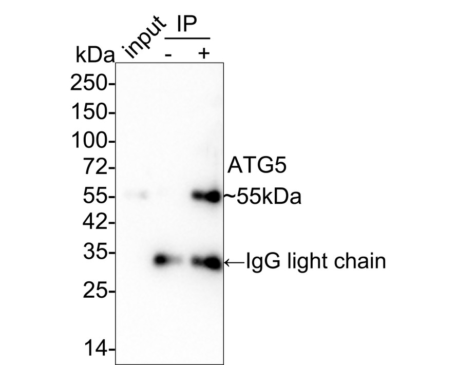
ATG5 was immunoprecipitated in 0.2mg HeLa cell lysate with ET1611-38 at 2 µg/25 µl agarose. Western blot was performed from the immunoprecipitate using ET1611-38 at 1/2,000 dilution. Anti-Rabbit IgG for IP Nano-secondary antibody (NBI01H) at 1/5,000 dilution was used for 1 hour at room temperature.
Lane 1: HeLa cell lysate (input)
Lane 2: Rabbit IgG instead of ET1611-38 in HeLa cell lysate
Lane 3: ET1611-38 IP in HeLa cell lysate
Blocking/Dilution buffer: 5% NFDM/TBST
Exposure time: 1 minute 21 seconds; ECL: K1802
请注意: All products are "FOR RESEARCH USE ONLY AND ARE NOT INTENDED FOR DIAGNOSTIC OR THERAPEUTIC USE"
LAPTM5 Confers the Resistance to Venetoclax via Promoting the Autophagosome-Lysosome Fusion in Multiple Myeloma
Author: Yuxiang Li, Jing Bai, Dan Liu, Jinxia Hao, Ruyu Yan, Hongjuan Guo, Yuzhi Huang, Hongtao Yu, Hao Leng, Kecheng Zhou, Minxia Liu
PMID: 39753521
期刊: Journal Of Cellular And Molecular Medicine
应用: WB
反应种属: Human
发表时间: 2025 Jan


Copyright © 广州杰特伟生物科技有限公司 All Rights Reserved. 备案号:粤ICP备19077843号