


p53 Recombinant Mouse Monoclonal Antibody [6B2-R]
Recombinant Mouse Monoclonal Antibody
Recombinant protein within C-terminal human p53.
Human, Monkey
WB, IF-Cell
Predicted band size: 53 kDa
293T cell lysate, HT-29 cell lysate, A431 cell lysate, COS-1 cell lysate, A431.
unconjugated
6B2-R
Liquid
1ug/ul
Store at +4℃ after thawing. Aliquot store at -20℃. Avoid repeated freeze / thaw cycles.
PBS (pH7.4), 0.1% BSA, 40% Glycerol. Preservative: 0.05% Sodium Azide.
IgG1
Protein A affinity purified.
WB
1:1,000-2,000
IF-Cell
1:100
Acts as a tumor suppressor in many tumor types; induces growth arrest or apoptosis depending on the physiological circumstances and cell type. Involved in cell cycle regulation as a trans-activator that acts to negatively regulate cell division by controlling a set of genes required for this process. One of the activated genes is an inhibitor of cyclin-dependent kinases. Apoptosis induction seems to be mediated either by stimulation of BAX and FAS antigen expression, or by repression of Bcl-2 expression. In cooperation with mitochondrial PPIF is involved in activating oxidative stress-induced necrosis; the function is largely independent of transcription. Induces the transcription of long intergenic non-coding RNA p21 (lincRNA-p21) and lincRNA-Mkln1. LincRNA-p21 participates in TP53-dependent transcriptional repression leading to apoptosis and seem to have to effect on cell-cycle regulation. Implicated in Notch signaling cross-over. Prevents CDK7 kinase activity when associated to CAK complex in response to DNA damage, thus stopping cell cycle progression. Isoform 2 enhances the transactivation activity of isoform 1 from some but not all TP53-inducible promoters. Isoform 4 suppresses transactivation activity and impairs growth suppression mediated by isoform 1. Isoform 7 inhibits isoform 1-mediated apoptosis. Regulates the circadian clock by repressing CLOCK-ARNTL/BMAL1-mediated transcriptional activation of PER2.
1. Louria-Hayon I et al. The promyelocytic leukemia protein protects p53 from Mdm2-mediated inhibition and degradation. J Biol Chem 278:33134-33141 (2003).
2. An W et al. Ordered cooperative functions of PRMT1, p300, and CARM1 in transcriptional activation by p53. Cell 117:735-748 (2004).
Nucleus. Cytoplasm. Localized in both nucleus and cytoplasm in most cells. In some cells, forms foci in the nucleus that are different from nucleoli.
Antigen NY-CO-13 antibody
BCC7 antibody
Cellular tumor antigen p53 antibody
FLJ92943 antibody
LFS1 antibody
Mutant tumor protein 53 antibody
p53 antibody
p53 tumor suppressor antibody
P53_HUMAN antibody
Phosphoprotein p53 antibody
展开
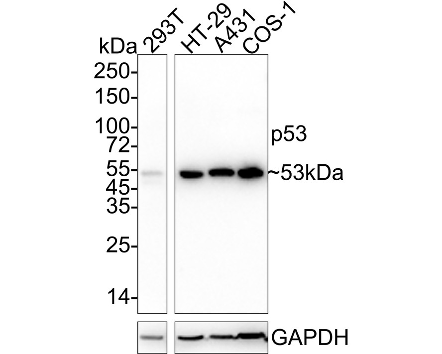
Western blot analysis of p53 on different lysates with Mouse anti-p53 antibody (HA601322) at 1/1,000 dilution.
Lane 1: 293T cell lysate
Lane 2: HT-29 cell lysate
Lane 3: A431 cell lysate
Lane 4: COS-1 cell lysate
Lysates/proteins at 20 µg/Lane.
Predicted band size: 53 kDa
Observed band size: 53 kDa
Exposure time: 46 seconds;
4-20% SDS-PAGE gel.
Proteins were transferred to a PVDF membrane and blocked with 5% NFDM/TBST for 1 hour at room temperature. The primary antibody (HA601322) at 1/1,000 dilution was used in 5% NFDM/TBST at 4℃ overnight. Goat Anti-Mouse IgG - HRP Secondary Antibody (HA1006) at 1/50,000 dilution was used for 1 hour at room temperature.
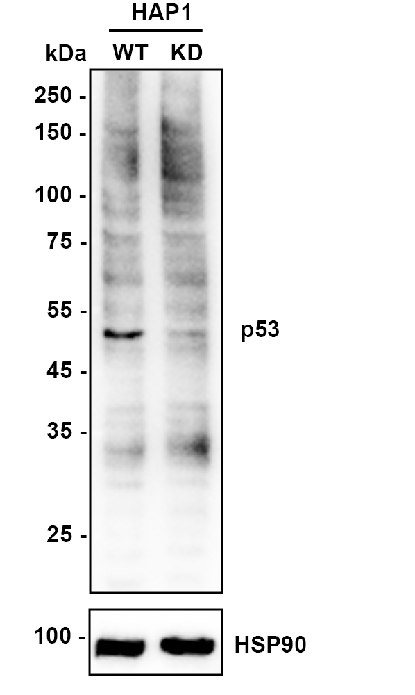
☑ Knockdown (KD)
Western blot analysis of p53 on different lysates with Mouse anti-p53 antibody (HA601322) at 1/2,000 dilution.
Lane 1: HAP1-parental cell lysate
Lane 2: HAP1-p53 KD cell lysate
Lysates/proteins at 10 µg/Lane.
Predicted band size: 53 kDa
Observed band size: 53 kDa
Exposure time: 80 seconds; ECL: K1801;
4-20% SDS-PAGE gel.
Proteins were transferred to a PVDF membrane and blocked with 5% NFDM/TBST for 1 hour at room temperature. The primary antibody (HA601322) at 1/2,000 dilution was used in K1803 at 4℃ overnight. Goat Anti-Mouse IgG - HRP Secondary Antibody (HA1006) at 1/50,000 dilution was used for 1 hour at room temperature.
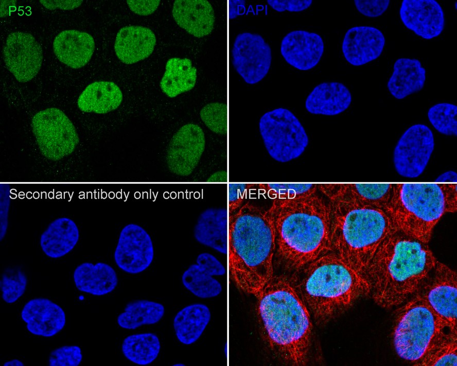
Immunocytochemistry analysis of A431 cells labeling p53 with Mouse anti-p53 antibody (HA601322) at 1/100 dilution.
Cells were fixed in 4% paraformaldehyde for 20 minutes at room temperature, permeabilized with 0.1% Triton X-100 in PBS for 5 minutes at room temperature, then blocked with 1% BSA in 10% negative goat serum for 1 hour at room temperature. Cells were then incubated with Mouse anti-p53 antibody (HA601322) at 1/100 dilution in 1% BSA in PBST overnight at 4 ℃. Goat Anti-Mouse IgG H&L (iFluor™ 488, HA1125) was used as the secondary antibody at 1/1,000 dilution. PBS instead of the primary antibody was used as the secondary antibody only control. Nuclear DNA was labelled in blue with DAPI.
beta Tubulin (ET1602-4, red) was stained at 1/100 dilution overnight at +4℃. Goat Anti-Rabbit IgG H&L (iFluor™ 594, HA1122) were used as the secondary antibody at 1/1,000 dilution.


Copyright © 广州杰特伟生物科技有限公司 All Rights Reserved. 备案号:粤ICP备19077843号