


NeuN Recombinant Rabbit Monoclonal Antibody [SR45-07]
Recombinant Rabbit monoclonal Antibody
Synthetic peptide within human NeuN aa 20-60.
Human, Mouse, Rat (Predicted: Cynomolgus monkey, Pig)
WB, IF-Cell, IF-Tissue, IHC-P, FC, IHC-Fr, mIHC
Predicted band size: 34 kDa
Mouse brain tissue lysate, rat brain tissue lysate, mouse cerebellum tissue lysate, rat cerebellum tissue lysate, SH-SY5Y cell lysate, SHG-44 cell lysate, rat cerebellum tissue, primary mouse neurons/glia cells, mouse cerebral cortex tissue, mouse cerebellum tissue, rat cerebral cortex tissue, mouse hippocampus tissue, rat hippocampus tissue, human brain tissue, human cerebellum tissue, human glioblastoma tissue, SH-SY5Y, mouse brain tissue.
unconjugated
SR45-07
Liquid
1ug/ul
Store at +4℃ after thawing. Aliquot store at -20℃ or -80℃. Avoid repeated freeze / thaw cycles.
1*TBS (pH7.4), 0.05% BSA, 40% Glycerol. Preservative: 0.05% Sodium Azide.
IgG
Protein A affinity purified.
WB
1:5,000-1:20,000
IF-Cell
1:250-1:500
IF-Tissue
1:500-1:1,000
IHC-P
1:200-1:10,000
IHC-Fr
1:1,000-1:2,000
FC
1:1,000
mIHC
1:1,000-1:10,000
| Mouse | 查看 12 篇文献如下 |
| Rat | 查看 8 篇文献如下 |
| mice | 查看 1 篇文献如下 |
Neuronal nuclei (NeuN, Fox-3, RBFOX3) is a nuclear protein expressed in most post-mitotic neurons of the central and peripheral nervous systems. NeuN is not detected in Purkinje cells, sympathetic ganglion cells, Cajal-Retzius cells, INL retinal cells, inferior olivary, and dentate nucleus neurons. This neuronal protein was originally identified by immunoreactivity with a monoclonal antibody also called NeuN. Using MS-analysis, NeuN was later identified as the Fox-3 gene product. Fox-3 contains an RNA recognition motif and functions as a splicing regulator. Fox-3 regulates alternative splicing of NumB, promoting neuronal differentiation during development.
1. Patel TP et al. Single-neuron NMDA receptor phenotype influences neuronal rewiring and reintegration following traumatic injury. J Neurosci 34:4200-13 (2014).
2. Kaur P et al. Expression profiling of RNA transcripts during neuronal maturation and ischemic injury. PLoS One 9:e103525 (2014).
Nucleus, Cytoplasm.
FLJ56884 antibody
FLJ58356 antibody
Fox-1 homolog C antibody
fox1 homolog C antibody
Fox3 antibody
FOX3NeuN antibody
hexaribonucleotide binding protein 3 antibody
HRNBP3 antibody
NEUN antibody
neuronal nuclei antibody
展开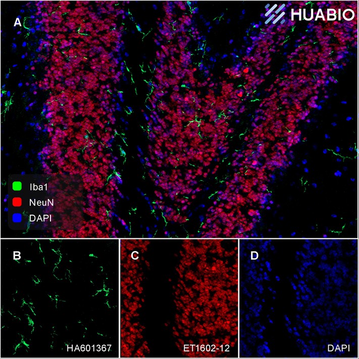
Application: IHC-Fr
Species: Mouse
Site: Hippocampus
Sample: Frozen section
Antibody concentration: 1:2,000
Antigen retrieval: Not required
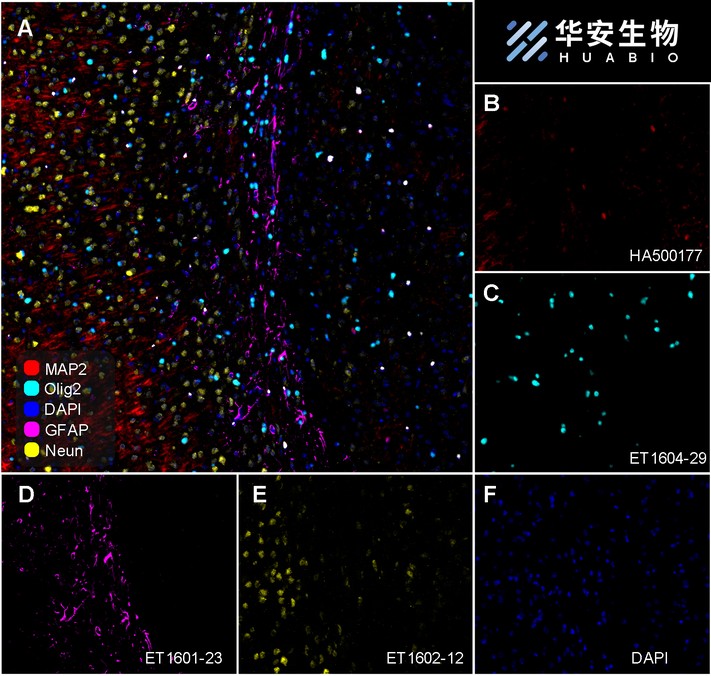
Fluorescence multiplex immunohistochemical analysis of mouse brain (Formalin/PFA-fixed paraffin-embedded sections). Panel A: the merged image of anti-MAP2 (HA500177, Red), anti-Olig2 (ET1604-29, Cyan), anti-GFAP (ET1601-23, Magenta) and anti-Neun (ET1602-12, Yellow) on mouse brain. HRP Conjugated UltraPolymer Goat Polyclonal Antibody HA1119/HA1120 was used as a secondary antibody. The immunostaining was performed with the Sequential Immuno-staining Kit (IRISKit™MH010101, www.luminiris.cn). The section was incubated in four rounds of staining: in the order of HA500177 (1/1,000 dilution), ET1604-29 (1/5,000 dilution), ET1601-23 (1/10,000 dilution) and ET1602-12 (1/10,000 dilution) for 20 mins at room temperature. Each round was followed by a separate fluorescent tyramide signal amplification system. Heat mediated antigen retrieval with Tris-EDTA buffer (pH 9.0) for 30 mins at 95℃. DAPI (blue) was used as a nuclear counter stain. Image acquisition was performed with Olympus VS200 Slide Scanner.
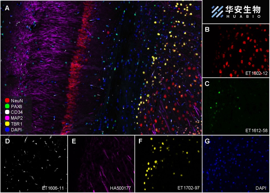
Fluorescence multiplex immunohistochemical analysis of mouse brain (Formalin/PFA-fixed paraffin-embedded sections). Panel A: the merged image of anti-NeuN (ET1602-12, red), anti-PAX6 (ET1612-58, green), anti-CD34 (ET1606-11, gray), anti-MAP2 (HA500177, magenta) and anti-TBR1 (ET1702-97, yellow) on mouse brain. HRP Conjugated UltraPolymer Goat Polyclonal Antibody HA1119/HA1120 was used as a secondary antibody. The immunostaining was performed with the Sequential Immuno-staining Kit (IRISKit™MH010101, www.luminiris.cn). The section was incubated in five rounds of staining: in the order of ET1602-12 (1/5,000 dilution), ET1612-58 (1/1,000 dilution), ET1606-11 (1/2,000 dilution), HA500177 (1/5,000 dilution) and ET1702-97 (1/1,000 dilution) for 20 mins at room temperature. Each round was followed by a separate fluorescent tyramide signal amplification system. Heat mediated antigen retrieval with Tris-EDTA buffer (pH 9.0) for 30 mins at 95℃. DAPI (blue) was used as a nuclear counter stain. Image acquisition was performed with Olympus VS200 Slide Scanner.
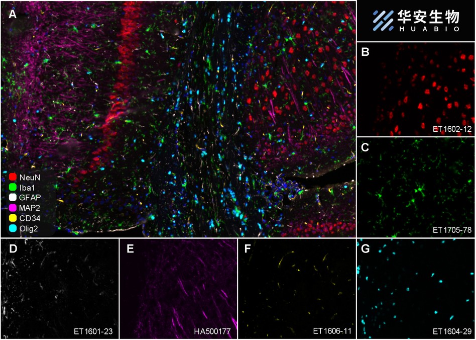
Fluorescence multiplex immunohistochemical analysis of mouse brain (Formalin/PFA-fixed paraffin-embedded sections). Panel A: the merged image of anti-NeuN (ET1602-12, red), anti-Iba1 (ET1705-78, green), anti-GFAP (ET1601-23, gray), anti-Olig2 (ET1604-29, cyan), anti-MAP2 (HA500177, magenta) and anti-CD34 (ET1606-11, yellow) on mouse brain. HRP Conjugated UltraPolymer Goat Polyclonal Antibody HA1119/HA1120 was used as a secondary antibody. The immunostaining was performed with the Sequential Immuno-staining Kit (IRISKit™MH010101, www.luminiris.cn). The section was incubated in six rounds of staining: in the order of ET1602-12(1/5,000 dilution), ET1705-78 (1/2,000 dilution), ET1601-23 (1/5,000 dilution), ET1604-29 (1/1,000 dilution), HA500177 (1/5,000 dilution) and ET1606-11 (1/2,000 dilution) for 20 mins at room temperature. Each round was followed by a separate fluorescent tyramide signal amplification system. Heat mediated antigen retrieval with Tris-EDTA buffer (pH 9.0) for 30 mins at 95℃. DAPI (blue) was used as a nuclear counter stain. Image acquisition was performed with Olympus VS200 Slide Scanner.
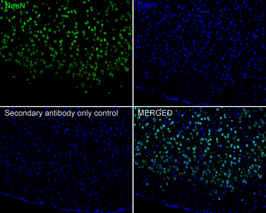
Application: IF-Tissue
Species: Human
Site: brain
Sample: Paraffin-embedded section
Antibody concentration: 1/500
Application: IF-tissue
Species: Mouse
Site: Cerebral cortex
Sample: Paraffin-embedded section
Antibody concentration: 1:1,000

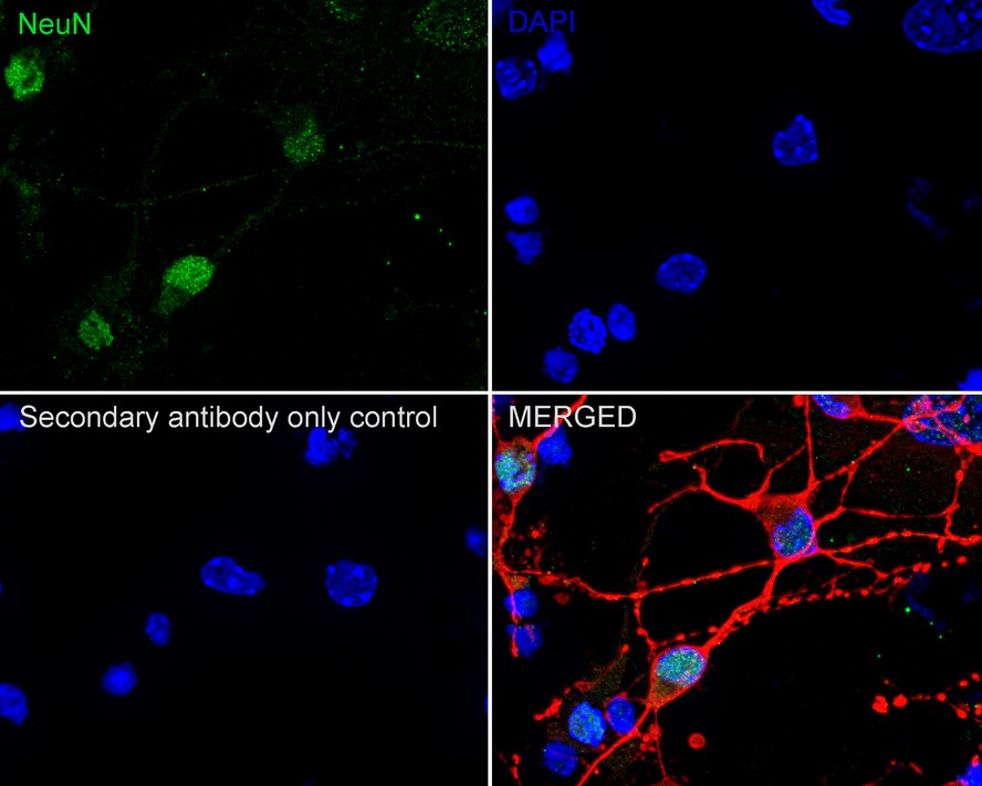
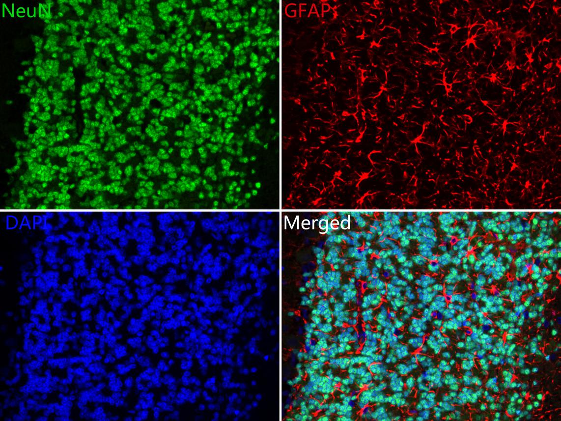
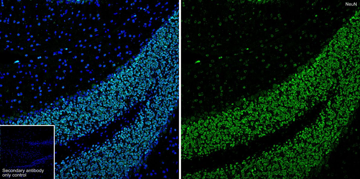
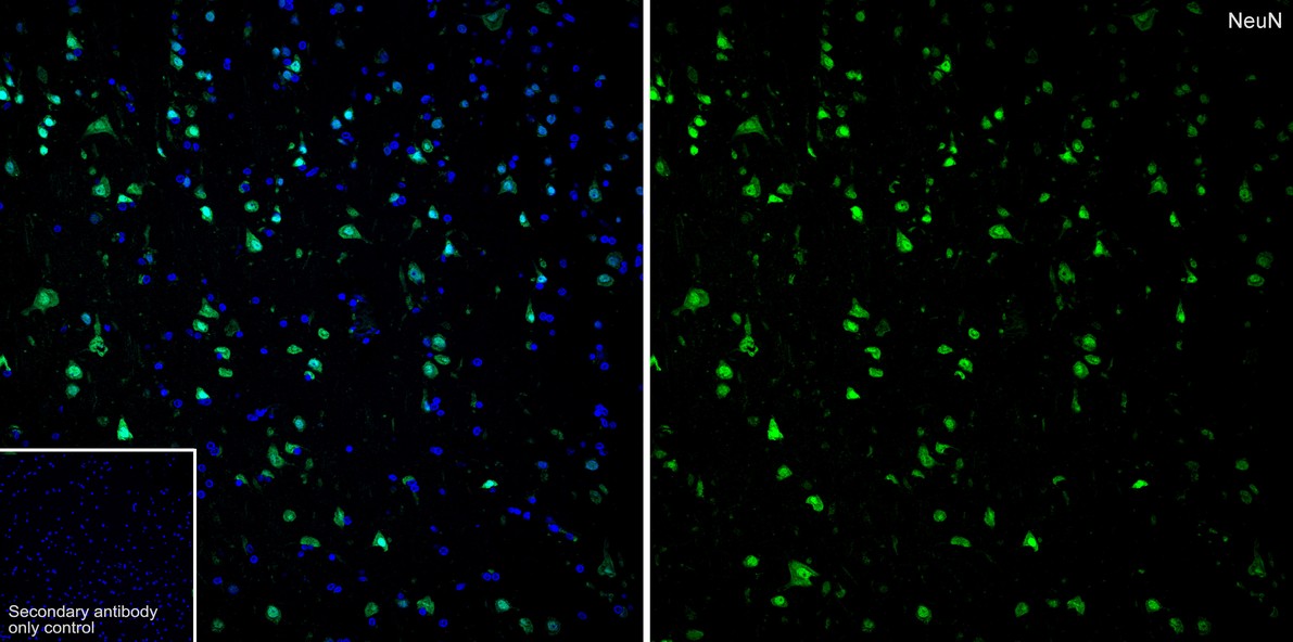
Immunocytochemistry analysis of primary mouse neurons/glia cells labeling NeuN with Rabbit anti-NeuN antibody (ET1602-12) at 1/500 dilution.
Cells were fixed with 4% PFA (15 min), permeabilized with 0.25% TritonX-100 for 15 minutes and then blocked with 1% BSA/10% normal goat serum/0.3M glycine in 0.1%PBS-Tween for 1h. The cells were then incubated overnight at 4℃ with Rabbit anti-NeuN antibody (ET1602-12) at at 1/500 dilution. Goat Anti-Rabbit IgG H&L (iFluor™ 488, HA1121) was used as the secondary antibody at 1/1,000 dilution. PBS instead of the primary antibody was used as the secondary antibody only control. Nuclear DNA was labelled in blue with DAPI.
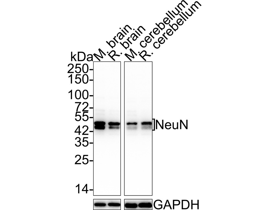
Western blot analysis of NeuN on different lysates with Rabbit anti-NeuN antibody (ET1602-12) at 1/5,000 dilution.
Lane 1: Mouse brain tissue lysate
Lane 2: Rat brain tissue lysate
Lane 3: Mouse cerebellum tissue lysate
Lane 4: Rat cerebellum tissue lysate
Lysates/proteins at 20 µg/Lane.
Predicted band size: 34 kDa
Observed band size: 45/50 kDa
Exposure time: 43 seconds;
4-20% SDS-PAGE gel.
Proteins were transferred to a PVDF membrane and blocked with 5% NFDM/TBST for 1 hour at room temperature. The primary antibody (ET1602-12) at 1/5,000 dilution was used in 5% NFDM/TBST at room temperature for 2 hours. Goat Anti-Rabbit IgG - HRP Secondary Antibody (HA1001) at 1/100,000 dilution was used for 1 hour at room temperature.

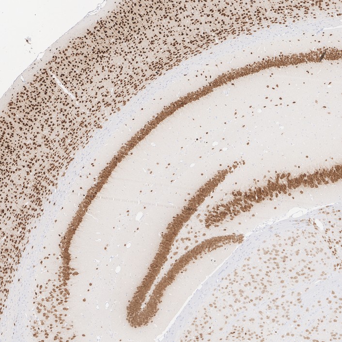
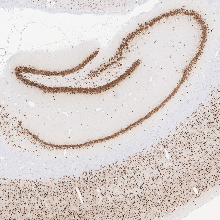
Immunohistochemical analysis of paraffin-embedded mouse brain tissue with Rabbit anti-NeuN antibody (ET1602-12) at 1/1,000 dilution.
The section was pre-treated using heat mediated antigen retrieval with sodium citrate buffer (pH 6.0) for 2 minutes. The tissues were blocked in 1% BSA for 20 minutes at room temperature, washed with ddH2O and PBS, and then probed with the primary antibody (ET1602-12) at 1/1,000 dilution for 1 hour at room temperature. The detection was performed using an HRP conjugated compact polymer system. DAB was used as the chromogen. Tissues were counterstained with hematoxylin and mounted with DPX.
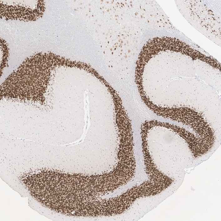
Immunohistochemical analysis of paraffin-embedded mouse cerebellum tissue with Rabbit anti-NeuN antibody (ET1602-12) at 1/1,000 dilution.
The section was pre-treated using heat mediated antigen retrieval with sodium citrate buffer (pH 6.0) for 2 minutes. The tissues were blocked in 1% BSA for 20 minutes at room temperature, washed with ddH2O and PBS, and then probed with the primary antibody (ET1602-12) at 1/1,000 dilution for 1 hour at room temperature. The detection was performed using an HRP conjugated compact polymer system. DAB was used as the chromogen. Tissues were counterstained with hematoxylin and mounted with DPX.
Immunohistochemical analysis of paraffin-embedded rat brain tissue with Rabbit anti-NeuN antibody (ET1602-12) at 1/1,000 dilution.
The section was pre-treated using heat mediated antigen retrieval with sodium citrate buffer (pH 6.0) for 2 minutes. The tissues were blocked in 1% BSA for 20 minutes at room temperature, washed with ddH2O and PBS, and then probed with the primary antibody (ET1602-12) at 1/1,000 dilution for 1 hour at room temperature. The detection was performed using an HRP conjugated compact polymer system. DAB was used as the chromogen. Tissues were counterstained with hematoxylin and mounted with DPX.
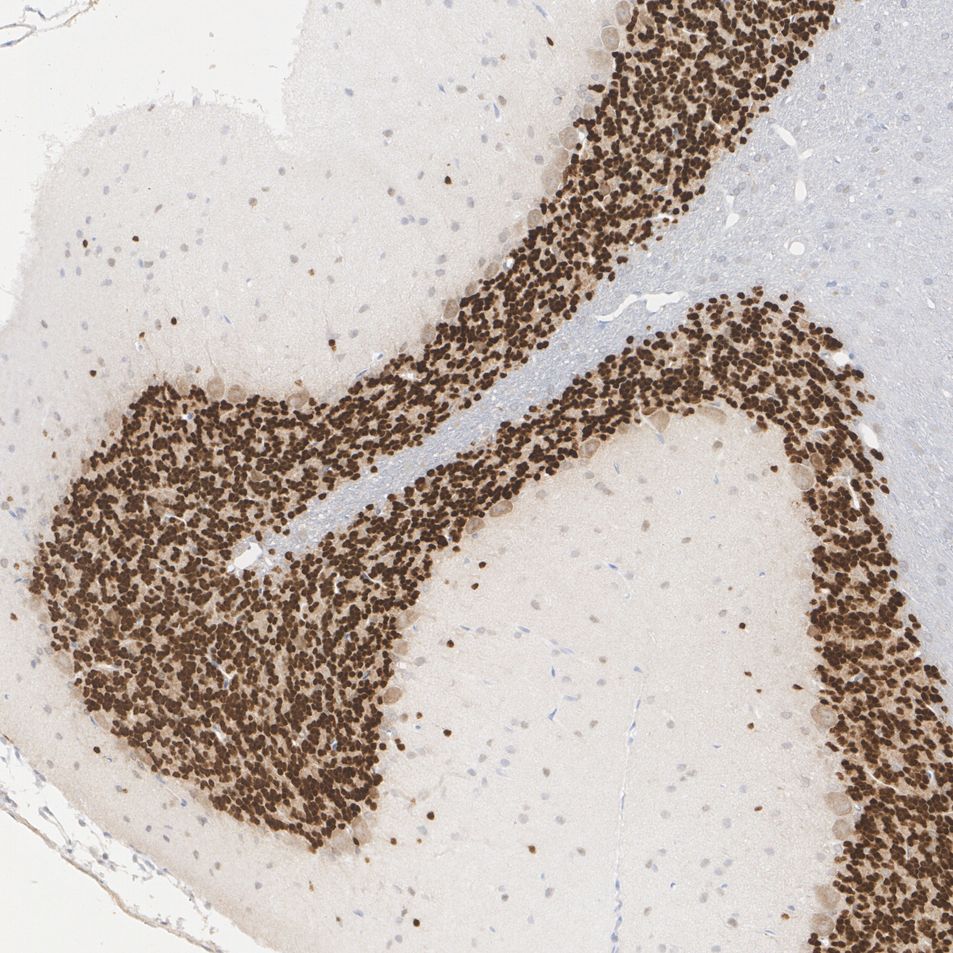
Immunohistochemical analysis of paraffin-embedded rat cerebellum tissue with Rabbit anti-NeuN antibody (ET1602-12) at 1/1,000 dilution.
The section was pre-treated using heat mediated antigen retrieval with sodium citrate buffer (pH 6.0) for 2 minutes. The tissues were blocked in 1% BSA for 20 minutes at room temperature, washed with ddH2O and PBS, and then probed with the primary antibody (ET1602-12) at 1/1,000 dilution for 1 hour at room temperature. The detection was performed using an HRP conjugated compact polymer system. DAB was used as the chromogen. Tissues were counterstained with hematoxylin and mounted with DPX.


Copyright © 广州杰特伟生物科技有限公司 All Rights Reserved. 备案号:粤ICP备19077843号