


STAT6 Recombinant Rabbit Monoclonal Antibody [SY13-09]
Recombinant Rabbit monoclonal Antibody
Synthetic peptide within human STAT6 C terminal.
Human, Mouse, Rat
WB, IF-Cell, IHC-P, IP, FC, IF-Tissue
Predicted band size: 94 kDa
Hela-si NT cell lysate, Hela-si STAT6 cell lysate, THP-1 cell lysate, Jurkat cell lysate, Raji cell lysate, HeLa cell lysate, MDA-MB-231 cell lysate, NIH/3T3 cell lysate, RAW264.7 cell lysate, human solitary fibrous tumor tissue, Hela, HepG2, NIH/3T3.
unconjugated
SY13-09
Liquid
1ug/ul
Store at +4℃ after thawing. Aliquot store at -20℃ or -80℃. Avoid repeated freeze / thaw cycles.
1*TBS (pH7.4), 0.05% BSA, 40% Glycerol. Preservative: 0.05% Sodium Azide.
IgG
Protein A affinity purified.
WB
1:2,000
IF-Cell
1:200-1:500
IHC-P
1:3,000
IF-Tissue
1:200
FC
1:1,000
IP
Use at an assay dependent concentration.
| Mouse | 查看 1 篇文献如下 |
Membrane receptor signaling by various ligands, including interferons and growth hormones such as EGF, induces activation of JAK kinases which then leads to tyrosine phosphorylation of proteins that have been designated Stats (signal transducers and activators of transcription). The first members of this family to be described include Stat1α p91, Stat1β p84 (a form of p91 that lacks 38 COOH-terminal amino acids) and Stat2 p113. Stat1 and Stat2 are induced by IFN-a and form a heterodimer which is part of the ISGF3 transcription factor complex. Stat3, which becomes activated in response to epidermal growth factor (EGF) and interleukin-6 (IL-6), but not interferon-γ (IFN-γ) or Stat4, is an additional member of this family. It has been suggested that the phosphorylated forms of both Stat3 and Stat4 form homodimers as well as heterodimers with the other members of the Stat family, and that differential activation of different Stat proteins in response to different ligands should help to explain specificity in nuclear signaling from the cell surface. Highest expression of Stat4 is seen in testis and myeloid cells. IL-12 has been identified as an activator of Stat4. Other members of the Stat family include Stat5, which has been shown to be activated by prolactin and by IL-3, and Stat6 (also designated IL-4 Stat), which is involved in IL-4-activated signaling pathways.
1. Zheng, C. et al. 2015. CD11b regulates obesity-induced insulin resistance via limiting alternative activation and proliferation of adipose tissue macrophages. Proc. Natl. Acad. Sci. U.S.A. 112: E7239-48.
2. Carlson, TJ. et al. 2014. Halofuginone-induced amino acid starvation regulates Stat3-dependent Th17 effector function and reduces established autoimmune inflammation. J. Immunol. 192: 2167-76.
Belongs to the transcription factor STAT family.
Tyrosine phosphorylated on Tyr-641 following stimulation by IL4/interleukin-4. Tyrosine phosphorylated following stimulation by IL3/interleukin-3 (By similarity). Dephosphorylation on tyrosine residues by PTPN2 negatively regulates the IL4/interleukin-4 mediated signaling.; Mono-ADP-ribosylated by PARP14.
Cytoplasm, Nucleus.
12S1644 antibody
D12S1644 antibody
IL 4 STAT antibody
IL-4 Stat antibody
IL4 STAT antibody
Interleukin 4 Induced antibody
Interleukin 4 Induced Transcription Factor IL4 STAT antibody
Signal transducer and activator of transcription 6 antibody
Signal Transducer And Activator Of Transcription 6 Interleukin 4 Induced antibody
Signal Transducer And Activator Of Transcription 6 Nirs Variant 1 antibody
展开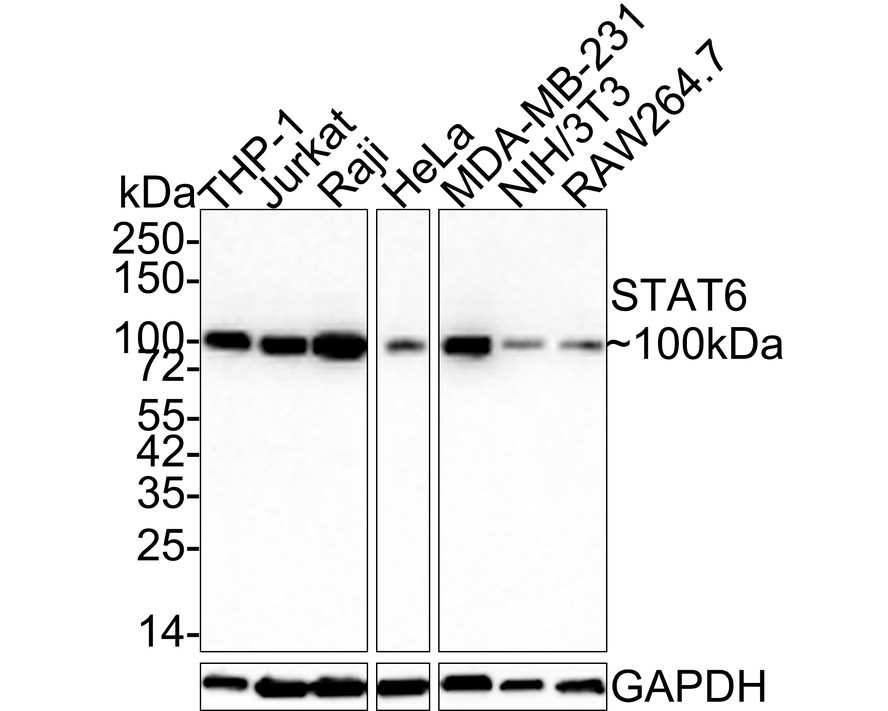
Western blot analysis of STAT6 on different lysates with Rabbit anti-STAT6 antibody (ET1605-49) at 1/2,000 dilution.
Lane 1: THP-1 cell lysate (20 µg/Lane)
Lane 2: Jurkat cell lysate (20 µg/Lane)
Lane 3: Raji cell lysate (20 µg/Lane)
Lane 4: HeLa cell lysate (20 µg/Lane)
Lane 5: MDA-MB-231 cell lysate (20 µg/Lane)
Lane 6: NIH/3T3 cell lysate (20 µg/Lane)
Lane 7: RAW264.7 cell lysate (20 µg/Lane)
Predicted band size: 94 kDa
Observed band size: 100 kDa
Exposure time: 2 minutes;
4-20% SDS-PAGE gel.
Proteins were transferred to a PVDF membrane and blocked with 5% NFDM/TBST for 1 hour at room temperature. The primary antibody (ET1605-49) at 1/2,000 dilution was used in 5% NFDM/TBST at room temperature for 2 hours. Goat Anti-Rabbit IgG - HRP Secondary Antibody (HA1001) at 1:100,000 dilution was used for 1 hour at room temperature.
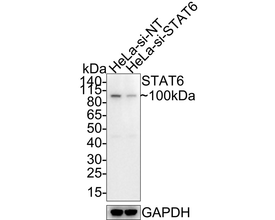
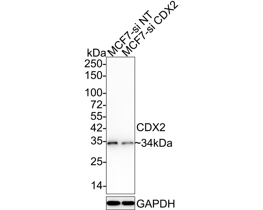
☑ Knockdown (KD)
Western blot analysis of STAT6 on different lysates with Rabbit anti-STAT6 antibody (ET1605-49) at 1/500 dilution.
Lane 1: Hela-si NT cell lysate
Lane 2: Hela-si STAT6 cell lysate
Lysates/proteins at 10 µg/Lane.
Predicted band size: 94 kDa
Observed band size: 100 kDa
Exposure time: 1 minute 34 seconds;
4-20% SDS-PAGE gel.
ET1605-49 was shown to specifically react with STAT6 in Hela-si NT cells. Weakened band was observed when Hela-si STAT6 sample was tested. Hela-si NT and Hela-si STAT6 samples were subjected to SDS-PAGE. Proteins were transferred to a PVDF membrane and blocked with 5% NFDM in TBST for 1 hour at room temperature. The primary antibody (ET1605-49, 1/500) and Loading control antibody (Rabbit anti-GAPDH, ET1601-4, 1/10,000) were used in 5% BSA at room temperature for 2 hours. Goat Anti-rabbit IgG-HRP Secondary Antibody (HA1001) at 1:300,000 dilution was used for 1 hour at room temperature.
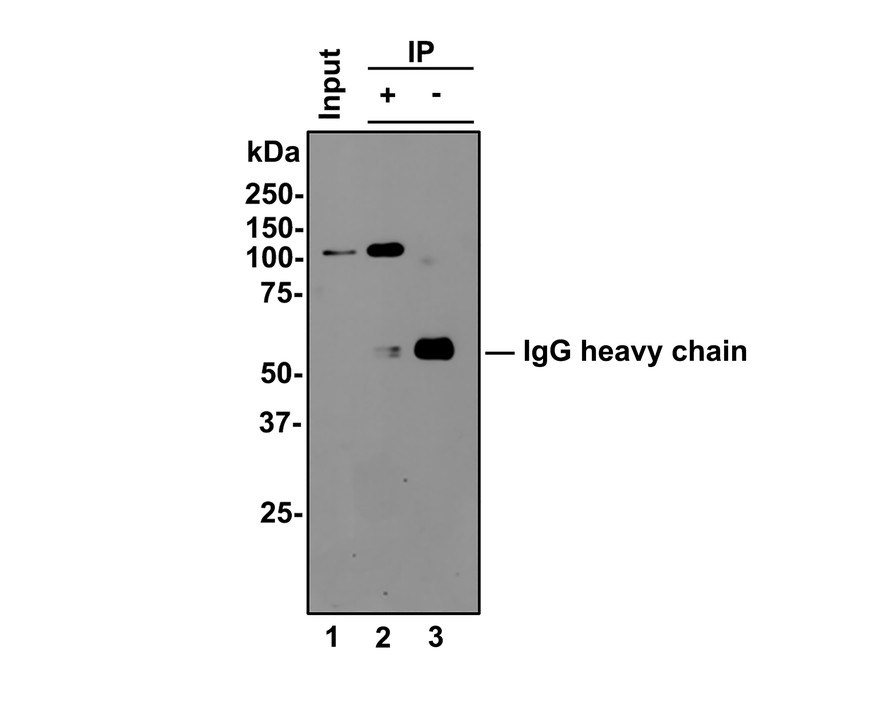
STAT6 was immunoprecipitated from 0.5 mg Hela whole cell lysates with ET1605-49 at 2 μg/mL. Western blot was performed from the immunoprecipitate using ET1605-49 at 1/500 dilution for 45 minutes at room temperature. Goat anti-Rabbit IgG-HRP Secondary Antibody (HA1001) was used at 1:300,000 dilution for 30 minutes at room temperature.
Lane 1: Hela whole cell lysates at 10 μg;
Lane 2: STAT6 (ET1605-49) IP in Hela whole cell lysates;
Lane 3: Rabbit IgG instead of STAT6 (ET1605-49) in Hela whole cell lysates.
Predicted band size: 94 kDa
Observed band size: 100 kDa
Exposure time: 2 minutes;
8% SDS-PAGE gel.
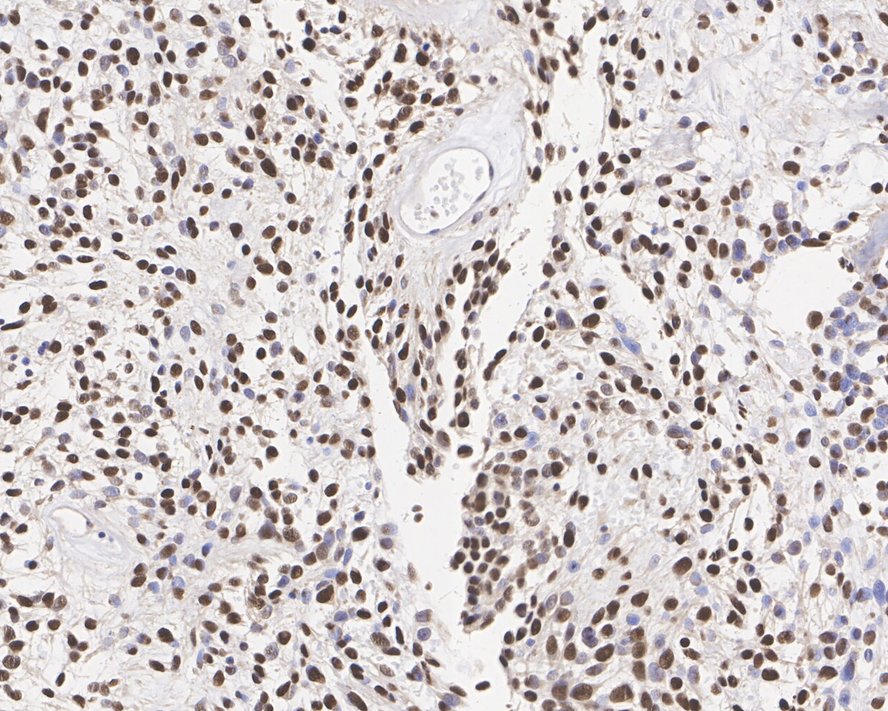
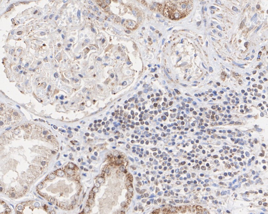
Immunohistochemical analysis of paraffin-embedded human solitary fibrous tumor tissue with Rabbit anti-STAT6 antibody (ET1605-49) at 1/1,000 dilution.
The section was pre-treated using heat mediated antigen retrieval with sodium citrate buffer (pH 6.0) for 2 minutes. The tissues were blocked in 1% BSA for 20 minutes at room temperature, washed with ddH2O and PBS, and then probed with the primary antibody (ET1605-49) at 1/1,000 dilution for 1 hour at room temperature. The detection was performed using an HRP conjugated compact polymer system. DAB was used as the chromogen. Tissues were counterstained with hematoxylin and mounted with DPX.
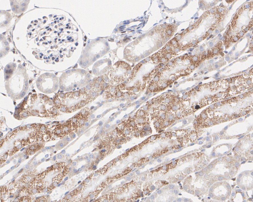
Immunohistochemical analysis of paraffin-embedded human kidney tissue with Rabbit anti-STAT6 antibody (ET1605-49) at 1/3,000 dilution.
The section was pre-treated using heat mediated antigen retrieval with sodium citrate buffer (pH 6.0) for 2 minutes. The tissues were blocked in 1% BSA for 20 minutes at room temperature, washed with ddH2O and PBS, and then probed with the primary antibody (ET1605-49) at 1/3,000 dilution for 1 hour at room temperature. The detection was performed using an HRP conjugated compact polymer system. DAB was used as the chromogen. Tissues were counterstained with hematoxylin and mounted with DPX.
Immunohistochemical analysis of paraffin-embedded mouse kidney tissue with Rabbit anti-STAT6 antibody (ET1605-49) at 1/3,000 dilution.
The section was pre-treated using heat mediated antigen retrieval with sodium citrate buffer (pH 6.0) for 2 minutes. The tissues were blocked in 1% BSA for 20 minutes at room temperature, washed with ddH2O and PBS, and then probed with the primary antibody (ET1605-49) at 1/3,000 dilution for 1 hour at room temperature. The detection was performed using an HRP conjugated compact polymer system. DAB was used as the chromogen. Tissues were counterstained with hematoxylin and mounted with DPX.
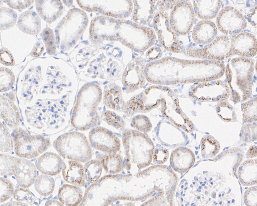
Immunohistochemical analysis of paraffin-embedded rat kidney tissue with Rabbit anti-STAT6 antibody (ET1605-49) at 1/3,000 dilution.
The section was pre-treated using heat mediated antigen retrieval with sodium citrate buffer (pH 6.0) for 2 minutes. The tissues were blocked in 1% BSA for 20 minutes at room temperature, washed with ddH2O and PBS, and then probed with the primary antibody (ET1605-49) at 1/3,000 dilution for 1 hour at room temperature. The detection was performed using an HRP conjugated compact polymer system. DAB was used as the chromogen. Tissues were counterstained with hematoxylin and mounted with DPX.
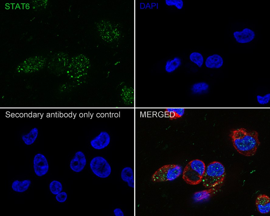
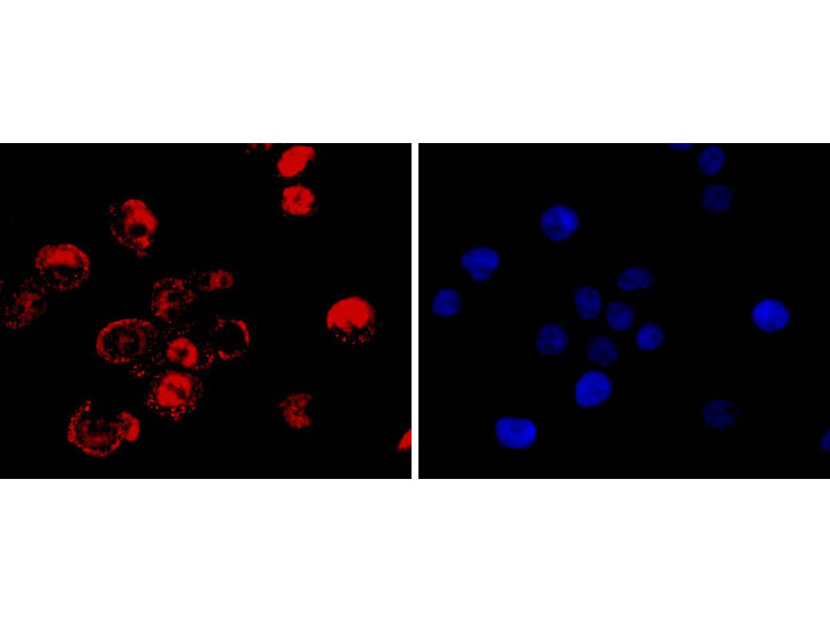
Immunocytochemistry analysis of MDA-MB-231 cells labeling STAT6 with Rabbit anti-STAT6 antibody (ET1605-49) at 1/500 dilution.
Cells were fixed in 4% paraformaldehyde for 20 minutes at room temperature, permeabilized with 0.1% Triton X-100 in PBS for 5 minutes at room temperature, then blocked with 1% BSA in 10% negative goat serum for 1 hour at room temperature. Cells were then incubated with Rabbit anti-STAT6 antibody (ET1605-49) at 1/500 dilution in 1% BSA in PBST overnight at 4 ℃. Goat Anti-Rabbit IgG H&L (iFluor™ 488, HA1121) was used as the secondary antibody at 1/1,000 dilution. PBS instead of the primary antibody was used as the secondary antibody only control. Nuclear DNA was labelled in blue with DAPI.
Beta tubulin (M1305-2, red) was stained at 1/100 dilution overnight at +4℃. Goat Anti-Mouse IgG H&L (iFluor™ 594, HA1126) was used as the secondary antibody at 1/1,000 dilution.
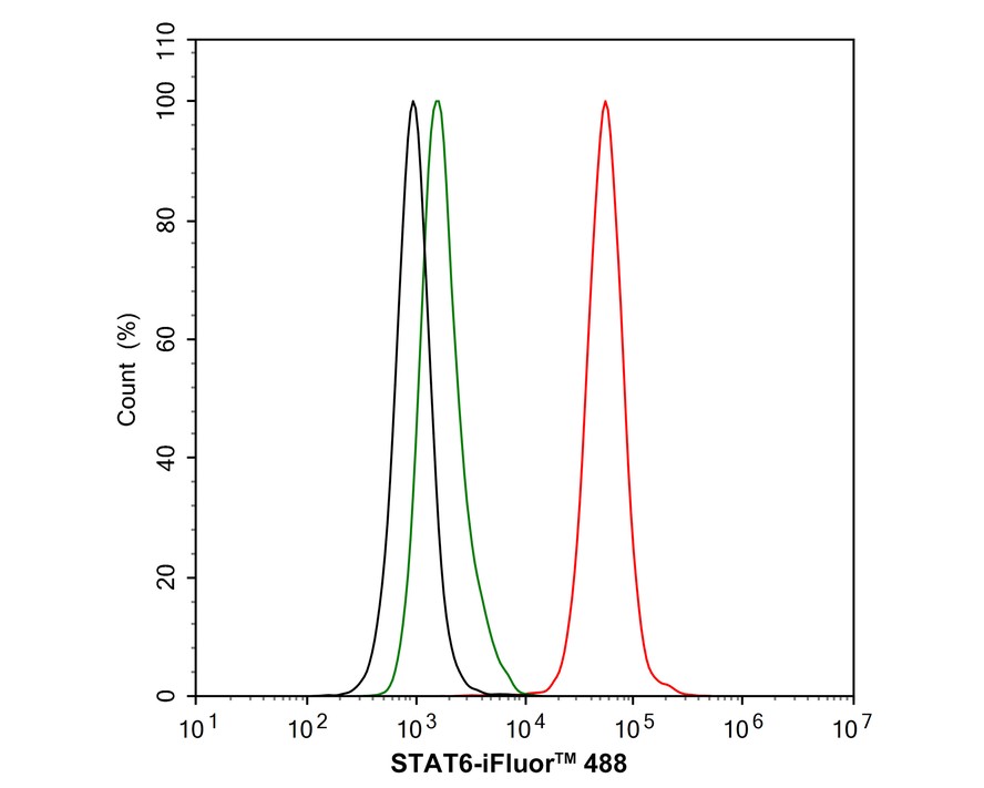
Flow cytometric analysis of HeLa cells labeling STAT6.
Cells were fixed and permeabilized. Then stained with the primary antibody (ET1605-49, 1μg/mL) (red) compared with Rabbit IgG Isotype Control (green). After incubation of the primary antibody at +4℃ for an hour, the cells were stained with a iFluor™ 488 conjugate-Goat anti-Rabbit IgG Secondary antibody (HA1121) at 1/1,000 dilution for 30 minutes at +4℃. Unlabelled sample was used as a control (cells without incubation with primary antibody; black).
Self-assembled PROTACs enable protein degradation to reprogram the tumor microenvironment for synergistically enhanced colorectal cancer immunotherapy
Author: Xinchen Lu,et al
PMID: 39386219
期刊: Bioactive Materials
应用: WB
反应种属: Mouse
发表时间: 2024 Oct


Copyright © 广州杰特伟生物科技有限公司 All Rights Reserved. 备案号:粤ICP备19077843号