


CDX2 Recombinant Rabbit Monoclonal Antibody [SY09-02]
Recombinant Rabbit monoclonal Antibody
Synthetic peptide within human CDX2 aa 20-60.
Human, Mouse, Rat
WB, IF-Cell, IF-Tissue, IHC-P, FC, CUT&Tag-seq
Predicted band size: 34 kDa
Caco-2 cell lysate, LoVo cell lysate, AGS cell lysate, LOVO, AGS, human appendix tissue, Human stomach cancer tissue, human colon cancer tissue, human colon tissue, mouse colon tissue, rat colon tissue, Hela.
unconjugated
SY09-02
Liquid
1ug/ul
Store at +4℃ after thawing. Aliquot store at -20℃ or -80℃. Avoid repeated freeze / thaw cycles.
1*TBS (pH7.4), 0.05% BSA, 40% Glycerol. Preservative: 0.05% Sodium Azide.
IgG
Protein A affinity purified.
WB
1:1,000-1:2,000
IF-Cell
1:50-1:200
IF-Tissue
1:200-1:2,000
IHC-P
1:500-20,000
FC
1:50-1:100
| IHC | 查看 1 篇文献如下 |
| Human | 查看 1 篇文献如下 |
In common with the two other Cdx genes, CDX2 regulates several essential processes in the development and function of the lower gastrointestinal tract (from the duodenum to the anus) in vertebrates. In vertebrate embryonic development, CDX2 becomes active in endodermal cells that are posterior to the developing stomach.These cells eventually form the intestinal epithelium. The activity of CDX2 at this stage is essential for the correct formation of the intestine and the anus.CDX2 is also required for the development of the placenta.
1. Yang Y et al. Heightened potency of human pluripotent stem cell lines created by transient BMP4 exposure. Proc Natl Acad Sci U S A 112:E2337-46 (2015).
2. Ohinata Y & Tsukiyama T Establishment of trophoblast stem cells under defined culture conditions in mice. PLoS One 9:e107308 (2014).
Belongs to the Caudal homeobox family.
Phosphorylation of Ser-60 mediates the transactivation capacity.
Nucleus.
Caudal type homeo box 2 antibody
Caudal type homeo box transcription factor 2 antibody
Caudal type homeobox 2 antibody
Caudal type homeobox protein 2 antibody
Caudal type homeobox transcription factor 2 antibody
Caudal-type homeobox protein 2 antibody
CDX 2 antibody
CDX 3 antibody
CDX-3 antibody
Cdx2 antibody
展开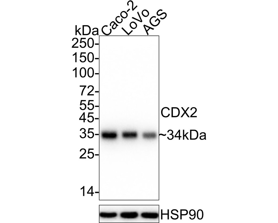
Western blot analysis of CDX2 on different lysates with Rabbit anti-CDX2 antibody (ET1605-4) at 1/1,000 dilution.
Lane 1: Caco-2 cell lysate
Lane 2: LoVo cell lysate
Lane 3: AGS cell lysate
Lysates/proteins at 15 µg/Lane.
Predicted band size: 34 kDa
Observed band size: 34 kDa
Exposure time: 25 seconds; ECL: K1801;
4-20% SDS-PAGE gel.
Proteins were transferred to a PVDF membrane and blocked with 5% NFDM/TBST for 1 hour at room temperature. The primary antibody (ET1605-4) at 1/1,000 dilution was used in 5% NFDM/TBST at 4℃ overnight. Goat Anti-Rabbit IgG - HRP Secondary Antibody (HA1001) at 1/50,000 dilution was used for 1 hour at room temperature.
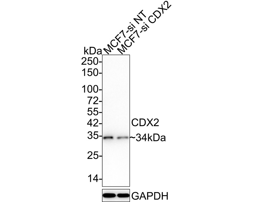
☑ Knockdown (KD)
Western blot analysis of CDX2 on different lysates with Rabbit anti-CDX2 antibody (ET1605-4) at 1/1,000 dilution.
Lane 1: MCF7-si NT cell lysate
Lane 2: MCF7-si CDX2 cell lysate
Lysates/proteins at 10 µg/Lane.
Predicted band size: 34 kDa
Observed band size: 34 kDa
Exposure time: 3 minutes;
4-20% SDS-PAGE gel.
Proteins were transferred to a PVDF membrane and blocked with 5% NFDM/TBST for 1 hour at room temperature. The primary antibody (ET1605-4) at 1/1,000 dilution was used in 5% NFDM/TBST at 4℃ overnight. Goat Anti-Rabbit IgG - HRP Secondary Antibody (HA1001) at 1/50,000 dilution was used for 1 hour at room temperature.
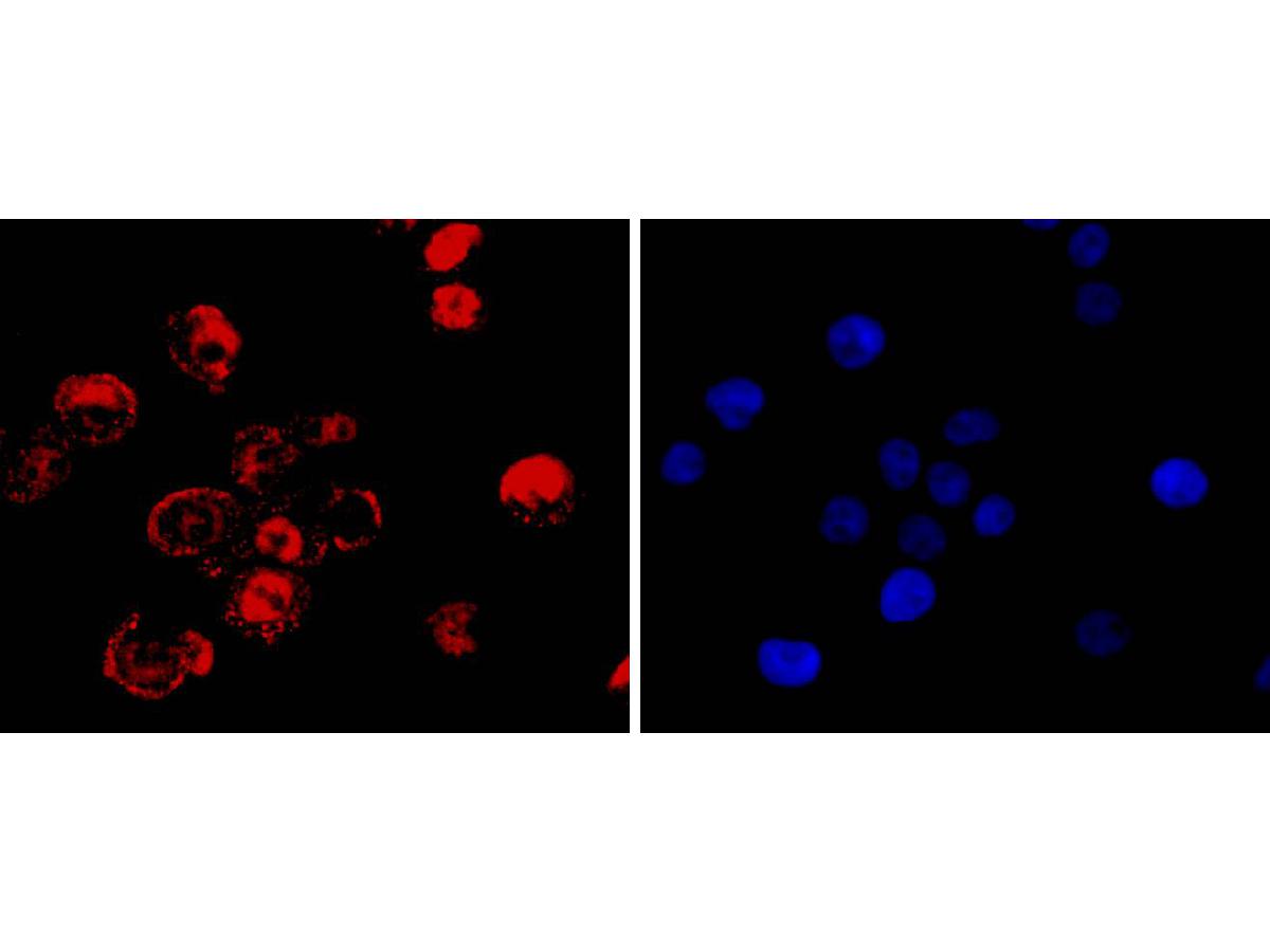
ICC staining of CDX2 in LOVO cells (red). Formalin fixed cells were permeabilized with 0.1% Triton X-100 in TBS for 10 minutes at room temperature and blocked with 1% Blocker BSA for 15 minutes at room temperature. Cells were probed with the primary antibody (ET1605-4, 1/50) for 1 hour at room temperature, washed with PBS. Alexa Fluor®488 Goat anti-Rabbit IgG was used as the secondary antibody at 1/1,000 dilution. The nuclear counter stain is DAPI (blue).
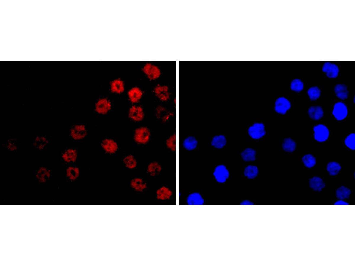
ICC staining of CDX2 in AGS cells (red). Formalin fixed cells were permeabilized with 0.1% Triton X-100 in TBS for 10 minutes at room temperature and blocked with 1% Blocker BSA for 15 minutes at room temperature. Cells were probed with the primary antibody (ET1605-4, 1/50) for 1 hour at room temperature, washed with PBS. Alexa Fluor®488 Goat anti-Rabbit IgG was used as the secondary antibody at 1/1,000 dilution. The nuclear counter stain is DAPI (blue).
Immunohistochemical analysis of paraffin-embedded human appendix tissue with Rabbit anti-CDX2 antibody (ET1605-4) at 1/5,000 dilution.
The section was pre-treated using heat mediated antigen retrieval with sodium citrate buffer (pH 6.0) for 2 minutes. The tissues were blocked in 1% BSA for 20 minutes at room temperature, washed with ddH2O and PBS, and then probed with the primary antibody (ET1605-4) at 1/5,000 dilution for 1 hour at room temperature. The detection was performed using an HRP conjugated compact polymer system. DAB was used as the chromogen. Tissues were counterstained with hematoxylin and mounted with DPX.
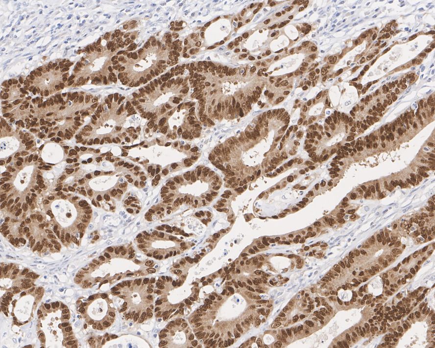
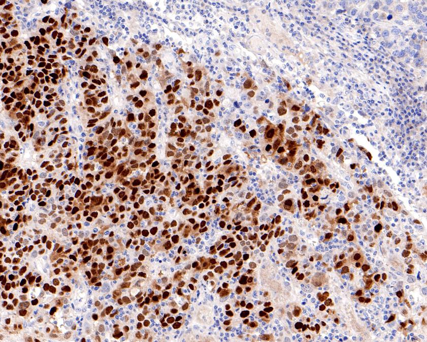
Immunohistochemical analysis of paraffin-embedded Human stomach cancer tissue with Rabbit anti-CDX2 antibody (ET1605-4) at 1/500 dilution.
The section was pre-treated using heat mediated antigen retrieval with sodium citrate buffer (pH 6.0) for 2 minutes. The tissues were blocked in 1% BSA for 20 minutes at room temperature, washed with ddH2O and PBS, and then probed with the primary antibody (ET1605-4) at 1/500 dilution for 1 hour at room temperature. The detection was performed using an HRP conjugated compact polymer system. DAB was used as the chromogen. Tissues were counterstained with hematoxylin and mounted with DPX.
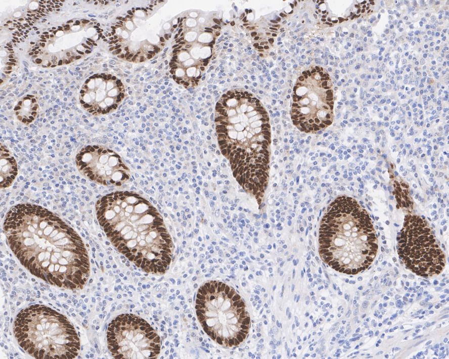
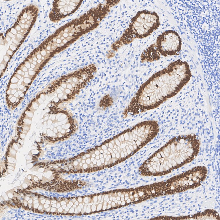
Immunohistochemical analysis of paraffin-embedded human colon cancer tissue with Rabbit anti-CDX2 antibody (ET1605-4) at 1/5,000 dilution.
The section was pre-treated using heat mediated antigen retrieval with sodium citrate buffer (pH 6.0) for 2 minutes. The tissues were blocked in 1% BSA for 20 minutes at room temperature, washed with ddH2O and PBS, and then probed with the primary antibody (ET1605-4) at 1/5,000 dilution for 1 hour at room temperature. The detection was performed using an HRP conjugated compact polymer system. DAB was used as the chromogen. Tissues were counterstained with hematoxylin and mounted with DPX.
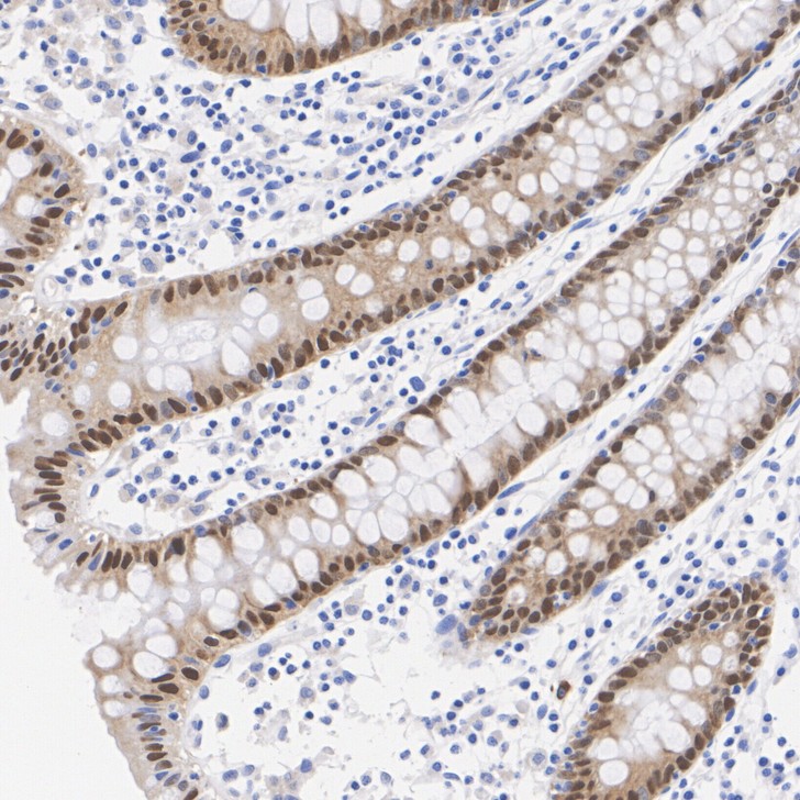
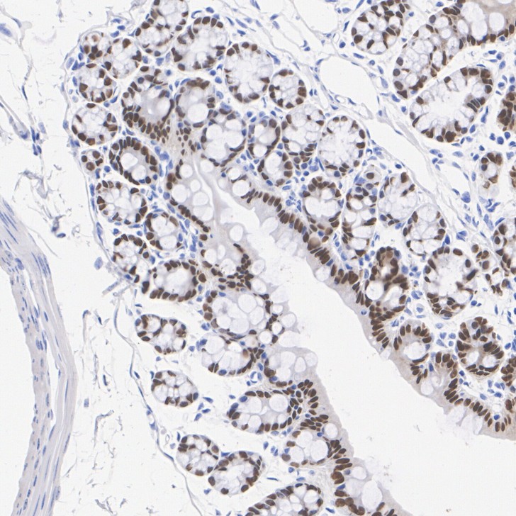
Immunohistochemical analysis of paraffin-embedded human colon tissue with Rabbit anti-CDX2 antibody (ET1605-4) at 1/20,000 dilution.
The section was pre-treated using heat mediated antigen retrieval with sodium citrate buffer (pH 6.0) for 2 minutes. The tissues were blocked in 1% BSA for 20 minutes at room temperature, washed with ddH2O and PBS, and then probed with the primary antibody (ET1605-4) at 1/20,000 dilution for 1 hour at room temperature. The detection was performed using an HRP conjugated compact polymer system. DAB was used as the chromogen. Tissues were counterstained with hematoxylin and mounted with DPX.
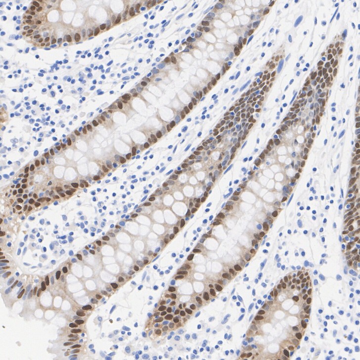
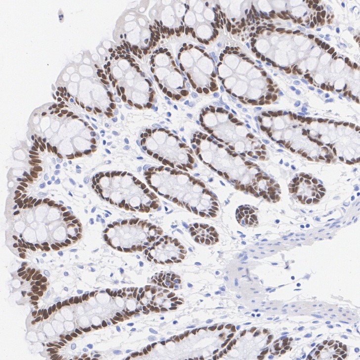
Immunohistochemical analysis of paraffin-embedded mouse colon tissue with Rabbit anti-CDX2 antibody (ET1605-4) at 1/20,000 dilution.
The section was pre-treated using heat mediated antigen retrieval with sodium citrate buffer (pH 6.0) for 2 minutes. The tissues were blocked in 1% BSA for 20 minutes at room temperature, washed with ddH2O and PBS, and then probed with the primary antibody (ET1605-4) at 1/20,000 dilution for 1 hour at room temperature. The detection was performed using an HRP conjugated compact polymer system. DAB was used as the chromogen. Tissues were counterstained with hematoxylin and mounted with DPX.
Immunohistochemical analysis of paraffin-embedded rat colon tissue with Rabbit anti-CDX2 antibody (ET1605-4) at 1/20,000 dilution.
The section was pre-treated using heat mediated antigen retrieval with sodium citrate buffer (pH 6.0) for 2 minutes. The tissues were blocked in 1% BSA for 20 minutes at room temperature, washed with ddH2O and PBS, and then probed with the primary antibody (ET1605-4) at 1/20,000 dilution for 1 hour at room temperature. The detection was performed using an HRP conjugated compact polymer system. DAB was used as the chromogen. Tissues were counterstained with hematoxylin and mounted with DPX.
☑ Relative expression (RE)
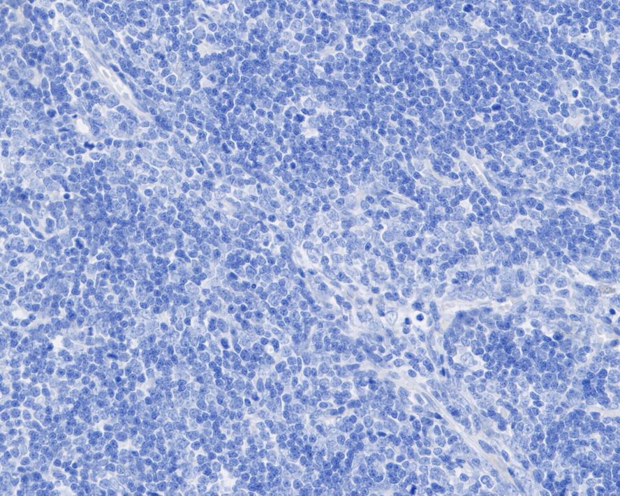
Immunohistochemical analysis of paraffin-embedded human thymus tissue (negative) with Rabbit anti-CDX2 antibody (ET1605-4) at 1/5,000 dilution.
The section was pre-treated using heat mediated antigen retrieval with sodium citrate buffer (pH 6.0) for 2 minutes. The tissues were blocked in 1% BSA for 20 minutes at room temperature, washed with ddH2O and PBS, and then probed with the primary antibody (ET1605-4) at 1/5,000 dilution for 1 hour at room temperature. The detection was performed using an HRP conjugated compact polymer system. DAB was used as the chromogen. Tissues were counterstained with hematoxylin and mounted with DPX.
☑ Relative expression (RE)
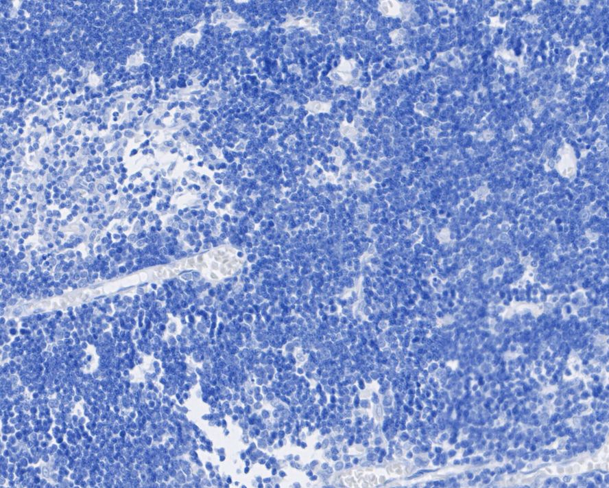
Immunohistochemical analysis of paraffin-embedded rat thymus tissue (negative) with Rabbit anti-CDX2 antibody (ET1605-4) at 1/5,000 dilution.
The section was pre-treated using heat mediated antigen retrieval with sodium citrate buffer (pH 6.0) for 2 minutes. The tissues were blocked in 1% BSA for 20 minutes at room temperature, washed with ddH2O and PBS, and then probed with the primary antibody (ET1605-4) at 1/5,000 dilution for 1 hour at room temperature. The detection was performed using an HRP conjugated compact polymer system. DAB was used as the chromogen. Tissues were counterstained with hematoxylin and mounted with DPX.
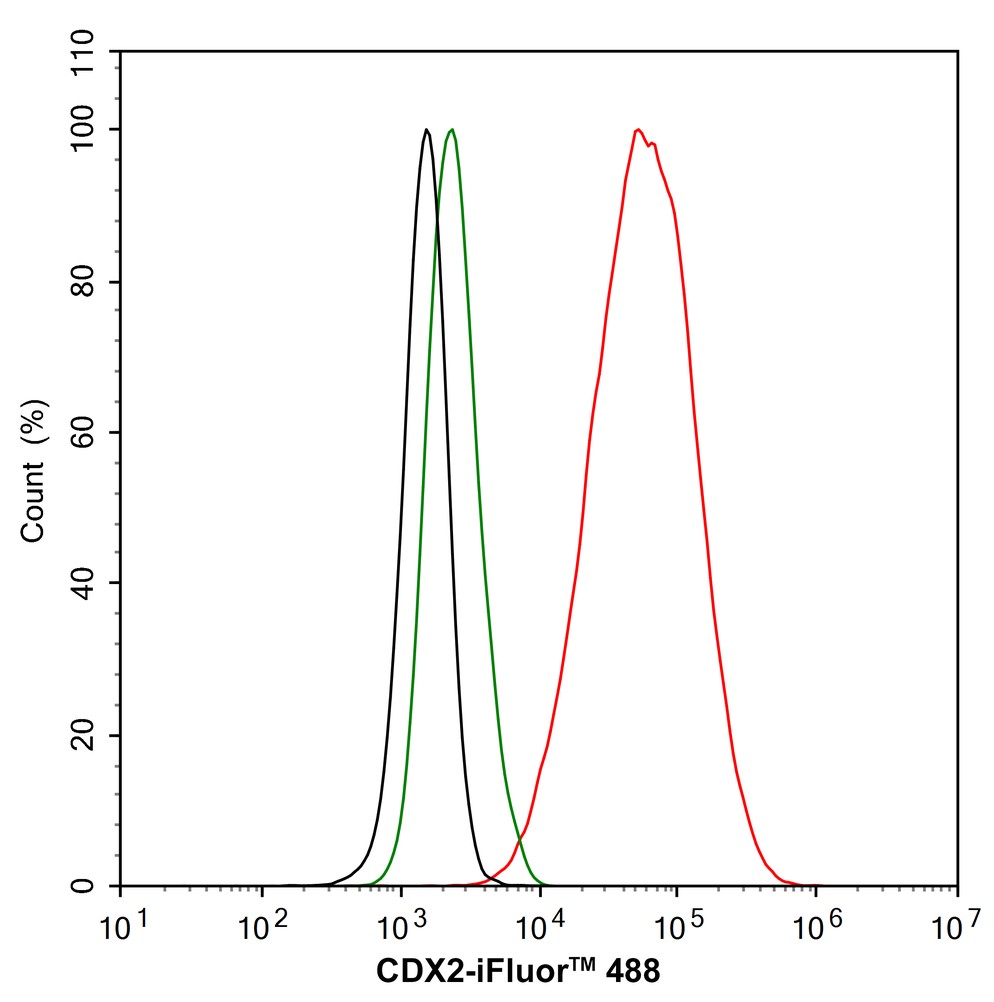
Flow cytometric analysis of CDX2 was done on Hela cells. The cells were fixed, permeabilized and stained with the primary antibody (ET1605-4, 1/50) (red). After incubation of the primary antibody at room temperature for an hour, the cells were stained with a Alexa Fluor 488-conjugated Goat anti-Rabbit IgG Secondary antibody at 1/1000 dilution for 30 minutes.Unlabelled sample was used as a control (cells without incubation with primary antibody; black).
Microfluidic droplet encapsulation‐guided organoid growth promotes parental tumor phenotype recapitulation
Author:
PMID: 37622267
期刊: International Journal Of Cancer
应用: IHC
反应种属: Human
发表时间: 2023 Aug


Copyright © 广州杰特伟生物科技有限公司 All Rights Reserved. 备案号:粤ICP备19077843号