Mouse PD-L1 Recombinant Rabbit Monoclonal Antibody [PSH05-03]
Recombinant Rabbit monoclonal Antibody
Recombinant protein within mouse PD-L1 aa 19-239 / 290.
Mouse
FC
Predicted band size: 33 kDa
RAW264.7 cells treated with 10μg/mL LPS for 8 hours.
unconjugated
PSH05-03
Liquid
1ug/ul
Store at +4℃ after thawing. Aliquot store at -20℃. Avoid repeated freeze / thaw cycles.
PBS (pH7.4).
IgG
Protein A affinity purified.
FC
1:1,000
Programmed death-ligand 1 (PD-L1) also known as cluster of differentiation 274 (CD274) or B7 homolog 1 (B7-H1) is a protein that in humans is encoded by the CD274 gene. Programmed death-ligand 1 (PD-L1) is a 40kDa type 1 transmembrane protein that has been speculated to play a major role in suppressing the adaptive arm of immune systems during particular events such as pregnancy, tissue allografts, autoimmune disease and other disease states such as hepatitis. Normally the adaptive immune system reacts to antigens that are associated with immune system activation by exogenous or endogenous danger signals. In turn, clonal expansion of antigen-specific CD8+ T cells and/or CD4+ helper cells is propagated. The binding of PD-L1 to the inhibitory checkpoint molecule PD-1 transmits an inhibitory signal based on interaction with phosphatases (SHP-1 or SHP-2) via Immunoreceptor Tyrosine-Based Switch Motif (ITSM). This reduces the proliferation of antigen-specific T-cells in lymph nodes, while simultaneously reducing apoptosis in regulatory T cells (anti-inflammatory, suppressive T cells) – further mediated by a lower regulation of the gene Bcl-2.
1. Lei Q et al. Resistance Mechanisms of Anti-PD1/PDL1 Therapy in Solid Tumors. Front Cell Dev Biol. 2020 Jul
2. Tran-Nguyen VK et al. Structure-based virtual screening for PDL1 dimerizers: Evaluating generic scoring functions. Curr Res Struct Biol. 2022 Jun
Cell membrane, Early endosome membrane, Recycling endosome membrane.
B7 H antibody
B7 H1 antibody
B7 homolog 1 antibody
B7-H1 antibody
B7H antibody
B7H1 antibody
CD 274 antibody
CD-274 antibody
CD274 antibody
CD274 antigen antibody
展开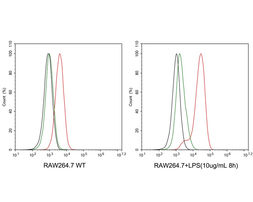
☑ Cell treatment (CT)
Flow cytometric analysis of RAW264.7 cells treated with 10μg/mL LPS for 8 hours labeling Mouse PD-L1.
Cells were washed twice with cold PBS and resuspend. Then stained with the primary antibody (HA722208, 1μg/mL) (red) compared with Rabbit IgG Isotype Control (green). After incubation of the primary antibody at +4℃ for an hour, the cells were stained with a iFluor™ 488 conjugate-Goat anti-Rabbit IgG Secondary antibody (HA1121) at 1/1,000 dilution for 30 minutes at +4℃. Unlabelled sample was used as a control (cells without incubation with primary antibody; black).


Huabio的研究人员会安排抗体进行不同的种属和应用验证,未列出的应用和种属是该产品不适用或者未经检测的。若您想在其它物种、应用中使用这个抗体产品,您可以通过邮件或者电话联系技术支持,获得更多抗体信息,为您的研究工作提供更多的帮助。


Copyright © 广州杰特伟生物科技有限公司 All Rights Reserved. 备案号:粤ICP备19077843号