FITC Conjugated Mouse PD-L1 Recombinant Rabbit Monoclonal Antibody [PSH05-04]
Recombinant Rabbit monoclonal Antibody
Recombinant protein within mouse PD-L1 aa 19-239 / 290.
Mouse
FC
Predicted band size: 33 kDa
RAW264.7 cells treated with 10μg/mL LPS for 8 hours.
FITC
PSH05-04
Liquid
1ug/ul
Store at 2℃ to 8℃. Avoid repeated freeze / thaw cycles.
Supplied in phosphate-buffered solution, pH 7.2, containing 0.2% ProClean 950 and BSA.
IgG
Protein A affinity purified.
FC
5 µl per million cells in 100 µl staining volume or 5 µl per 100 µl of whole blood.
Programmed death-ligand 1 (PD-L1) also known as cluster of differentiation 274 (CD274) or B7 homolog 1 (B7-H1) is a protein that in humans is encoded by the CD274 gene. Programmed death-ligand 1 (PD-L1) is a 40kDa type 1 transmembrane protein that has been speculated to play a major role in suppressing the adaptive arm of immune systems during particular events such as pregnancy, tissue allografts, autoimmune disease and other disease states such as hepatitis. Normally the adaptive immune system reacts to antigens that are associated with immune system activation by exogenous or endogenous danger signals. In turn, clonal expansion of antigen-specific CD8+ T cells and/or CD4+ helper cells is propagated. The binding of PD-L1 to the inhibitory checkpoint molecule PD-1 transmits an inhibitory signal based on interaction with phosphatases (SHP-1 or SHP-2) via Immunoreceptor Tyrosine-Based Switch Motif (ITSM). This reduces the proliferation of antigen-specific T-cells in lymph nodes, while simultaneously reducing apoptosis in regulatory T cells (anti-inflammatory, suppressive T cells) – further mediated by a lower regulation of the gene Bcl-2.
1. Lei Q et al. Resistance Mechanisms of Anti-PD1/PDL1 Therapy in Solid Tumors. Front Cell Dev Biol. 2020 Jul
2. Tran-Nguyen VK et al. Structure-based virtual screening for PDL1 dimerizers: Evaluating generic scoring functions. Curr Res Struct Biol. 2022 Jun
Cell membrane, Early endosome membrane, Recycling endosome membrane.
B7 H antibody
B7 H1 antibody
B7 homolog 1 antibody
B7-H1 antibody
B7H antibody
B7H1 antibody
CD 274 antibody
CD-274 antibody
CD274 antibody
CD274 antigen antibody
展开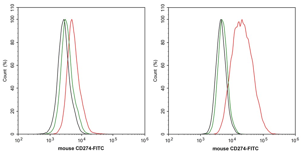
☑ Cell treatment (CT)
Flow cytometric analysis of RAW264.7 cells treated with or without 10μg/mL LPS for 8 hours labeling Mouse PD-L1.
Cells were washed twice with cold PBS and resuspend. Then incubated for 1 hour at +4℃ with Mouse PD-L1 (HA720213F, red, 1μg/mL) and Rabbit IgG Isotype Control (FITC, green, 1μg/mL). Unlabelled sample was used as a control (cells without incubation with primary antibody; black).


Copyright © 广州杰特伟生物科技有限公司 All Rights Reserved. 备案号:粤ICP备19077843号