Human CD5 Recombinant Rabbit Monoclonal Antibody [PSH05-01]
Recombinant Rabbit monoclonal Antibody
Recombinant protein within human CD5 aa 25-372 / 495.
Human
IF-Cell, FC
Predicted band size: 55 kDa
Jurkat, human peripheral blood lymphocytes
unconjugated
PSH05-01
Liquid
1ug/ul
Store at +4℃ after thawing. Aliquot store at -20℃. Avoid repeated freeze / thaw cycles.
PBS (pH7.4), 0.1% BSA, 40% Glycerol. Preservative: 0.05% Sodium Azide.
IgG
Protein A affinity purified.
IF-Cell
1:100
FC
1:1,000
CD5 is a cluster of differentiation expressed on the surface of T cells (various species) and in a subset of murine B cells known as B-1a. The expression of this receptor in human B cells has been a controversial topic and to date there is no consensus regarding the role of this receptor as a marker of human B cells. B-1 cells have limited diversity of their B-cell receptor due to their lack of the enzyme terminal deoxynucleotidyl transferase (TdT) and are potentially self-reactive. CD5 serves to mitigate activating signals from the BCR so that the B-1 cells can only be activated by very strong stimuli (such as bacterial proteins) and not by normal tissue proteins. CD5 was used as a T-cell marker until monoclonal antibodies against CD3 were developed. In humans, the gene is located on the long arm of chromosome 11. There is no confirmed ligand for CD5 but there is evidence that CD72, a C-type lectin, may be a ligand or that CD5 may be homophilic, binding CD5 on the surface of other cells. CD5 includes a scavenger receptor cysteine-rich protein domain. T cells express higher levels of CD5 than B cells. CD5 is upregulated on T cells upon strong activation. In the thymus, there is a correlation with CD5 expression and strength of the interaction of the T cell towards self-peptides.
1. He M et al. CD5 expression by dendritic cells directs T cell immunity and sustains immunotherapy responses. Science. 2023 Feb
2. Durani U et al. CD5+ diffuse large B-cell lymphoma: a narrative review. Leuk Lymphoma. 2021 Dec
Cell membrane.
CD 5 antibody
CD5 antibody
CD5 antigen (p56 62) antibody
CD5 antigen antibody
CD5 molecule antibody
CD5_HUMAN antibody
LEU1 antibody
LyA antibody
Lymphocyte antigen T1/Leu-1 antibody
OTTHUMP00000236973 antibody
展开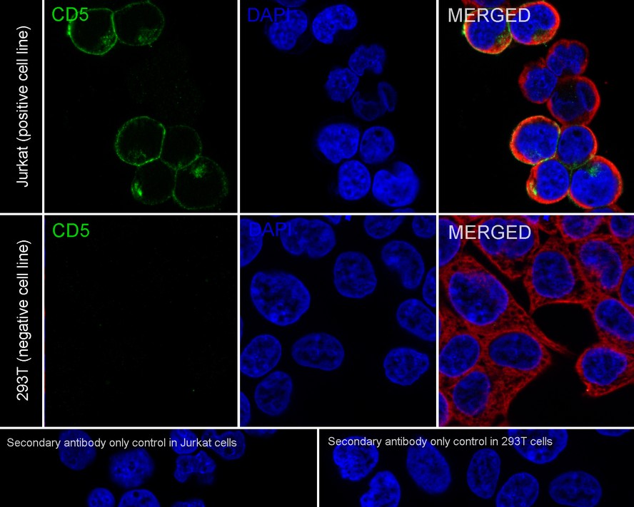
☑ Relative expression (RE)
Immunocytochemistry analysis of Jurkat (positive) and 293T (negative) labeling Human CD5 with Rabbit anti-Human CD5 antibody (HA722210) at 1/100 dilution.
Cells were fixed in 4% paraformaldehyde for 20 minutes at room temperature, permeabilized with 0.1% Triton X-100 in PBS for 5 minutes at room temperature, then blocked with 1% BSA in 10% negative goat serum for 1 hour at room temperature. Cells were then incubated with Rabbit anti-Human CD5 antibody (HA722210) at 1/100 dilution in 1% BSA in PBST overnight at 4 ℃. Goat Anti-Rabbit IgG H&L (iFluor™ 488, HA1121) was used as the secondary antibody at 1/1,000 dilution. PBS instead of the primary antibody was used as the secondary antibody only control. Nuclear DNA was labelled in blue with DAPI.
Beta tubulin (M1305-2, red) was stained at 1/100 dilution overnight at +4℃. Goat Anti-Mouse IgG H&L (iFluor™ 594, HA1126) was used as the secondary antibody at 1/1,000 dilution.
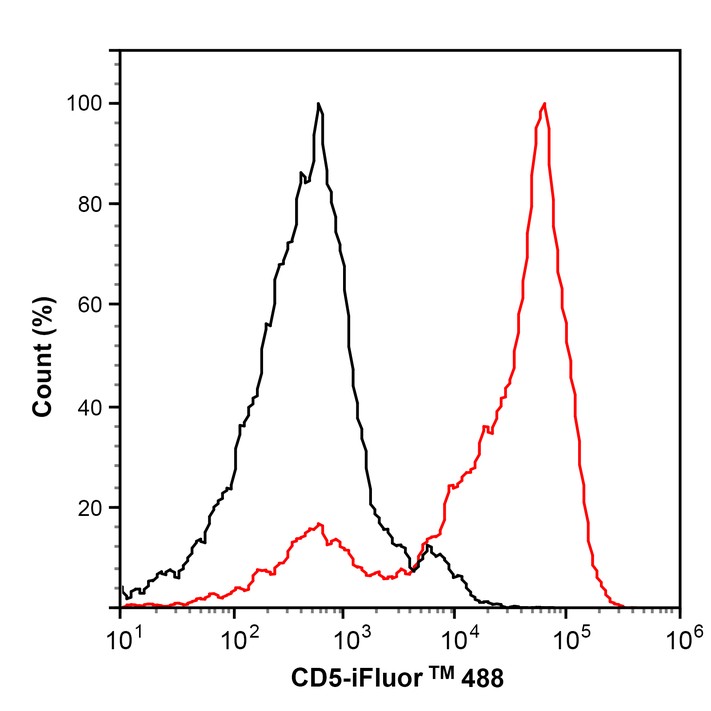
Flow cytometric analysis of human peripheral blood lymphocytes labeling Human CD5.
Cells were washed twice with cold PBS and resuspend. Then stained with the primary antibody (HA722210, 1μg/mL) (red). After incubation of the primary antibody at +4℃ for an hour, the cells were stained with a iFluor™ 488 conjugate-Goat anti-Rabbit IgG Secondary antibody (HA1121) at 1/1,000 dilution for 30 minutes at +4℃. Unlabelled sample was used as a control (cells without incubation with primary antibody; black).


Copyright © 广州杰特伟生物科技有限公司 All Rights Reserved. 备案号:粤ICP备19077843号