


HER3 Recombinant Rabbit Monoclonal Antibody [R5]
Recombinant Rabbit monoclonal Antibody
Recombinant protein within Human HER3 Extracellular domain.
Human
FC
Predicted band size: 148 kDa
MCF-7, SK-Br-3.
unconjugated
R5
Liquid
1ug/ul
Store at +4℃ after thawing. Aliquot store at -20℃. Avoid repeated freeze / thaw cycles.
PBS (pH7.4), 0.05% BSA, 40% Glycerol. Preservative: 0.05% Sodium Azide.
IgG
Protein A affinity purified.
FC
1:100-1:1,000
This gene encodes a member of the epidermal growth factor receptor (EGFR) family of receptor tyrosine kinases. This membrane-bound protein has a neuregulin binding domain but not an active kinase domain. It therefore can bind this ligand but not convey the signal into the cell through protein phosphorylation. However, it does form heterodimers with other EGF receptor family members which do have kinase activity. Heterodimerization leads to the activation of pathways which lead to cell proliferation or differentiation. Amplification of this gene and/or overexpression of its protein have been reported in numerous cancers, including prostate, bladder, and breast tumors. Alternate transcriptional splice variants encoding different isoforms have been characterized. One isoform lacks the intermembrane region and is secreted outside the cell. This form acts to modulate the activity of the membrane-bound form. Additional splice variants have also been reported, but they have not been thoroughly characterized.
1. Koganemaru S. et. al. U3-1402, a Novel HER3-Targeting Antibody-Drug Conjugate, for the Treatment of Colorectal Cancer. Mol Cancer Ther. 2019 Nov
2. Hyman DM. et. al. HER kinase inhibition in patients with HER2- and HER3-mutant cancers. Nature. 2018 Feb
Cell membrane; Secreted.
c erbB 3 antibody
c erbB3 antibody
Erb b2 receptor tyrosine kinase 3 antibody
ErbB 3 antibody
ERBB3 antibody
ERBB3 protein antibody
erbB3 S antibody
ERBB3_HUMAN antibody
Glial growth factor receptor antibody
HER 3 antibody
展开
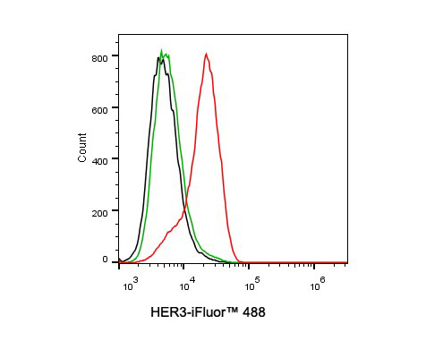
Flow cytometric analysis of MCF-7 cells labeling HER3.
Cells were washed twice with cold PBS and resuspend. Then stained with the primary antibody (HA721126, 0.1ug/ml) (red) compared with Rabbit IgG Isotype Control (green). After incubation of the primary antibody at +4℃ for an hour, the cells were stained with a iFluor™ 488 conjugate-Goat anti-Rabbit IgG Secondary antibody (HA1121) at 1/1,000 dilution for 30 minutes at +4℃. Unlabelled sample was used as a control (cells without incubation with primary antibody; black).

Flow cytometric analysis of SK-Br-3 cells labeling HER3.
Cells were washed twice with cold PBS and resuspend. Then stained with the primary antibody (HA721126, 1ug/ml) (red) compared with Rabbit IgG Isotype Control (green). After incubation of the primary antibody at +4℃ for an hour, the cells were stained with a iFluor™ 488 conjugate-Goat anti-Rabbit IgG Secondary antibody (HA1121) at 1/1,000 dilution for 30 minutes at +4℃. Unlabelled sample was used as a control (cells without incubation with primary antibody; black).


Copyright © 广州杰特伟生物科技有限公司 All Rights Reserved. 备案号:粤ICP备19077843号