


Myc tag Rabbit Polyclonal Antibody
Rabbit Polyclonal Antibody
Synthetic peptide within human Myc aa 410-420.
Species independent
WB, IP, IF-Cell, FC
PG-CM cell lysates, C-terminal Myc-tagged recombinant protein, N-terminal Myc-tagged recombinant protein.
unconjugated
Liquid
1ug/ul
Store at +4℃ after thawing. Aliquot store at -20℃ or -80℃. Avoid repeated freeze / thaw cycles.
1*PBS (pH7.4), 0.2% BSA, 40% Glycerol. Preservative: 0.05% Sodium Azide.
IgG
Immunogen affinity purified.
WB
1:20,000-1:50,000
IP
2-5 µg/ml.
IF-Cell
1:200
FC
1:1,000
| WB | 查看 60 篇文献如下 |
| IP | 查看 11 篇文献如下 |
| CoIP | 查看 5 篇文献如下 |
| IF | 查看 5 篇文献如下 |
| Co-IP | 查看 2 篇文献如下 |
| ICC | 查看 1 篇文献如下 |
| CO-IP | 查看 1 篇文献如下 |
| IB | 查看 1 篇文献如下 |
| Human | 查看 39 篇文献如下 |
| Chicken | 查看 4 篇文献如下 |
| Mouse | 查看 4 篇文献如下 |
| Pig | 查看 3 篇文献如下 |
| Fish | 查看 2 篇文献如下 |
| 293T cells | 查看 1 篇文献如下 |
| HEK293 cells | 查看 1 篇文献如下 |
| Xanthomonas oryzae pv. oryzicola | 查看 1 篇文献如下 |
| Circovirus | 查看 1 篇文献如下 |
| zebrafish | 查看 1 篇文献如下 |
| pig | 查看 1 篇文献如下 |
| Porcine Circovirus | 查看 1 篇文献如下 |
| Bombyx Mori | 查看 1 篇文献如下 |
| Zebrafish | 查看 1 篇文献如下 |
| Kiwifruit | 查看 1 篇文献如下 |
| Persimmon fruit | 查看 1 篇文献如下 |
| Nicotiana benthamiana | 查看 1 篇文献如下 |
| Escherichia coli | 查看 1 篇文献如下 |
| Dog | 查看 1 篇文献如下 |
Myc gene encodes for a transcription factor that is believed to regulate expression of 15% of all genes through binding on Enhancer Box sequences (E-boxes) and recruiting histone acetyltransferases (HATs). c-Myc is commonly activated in a variety of tumor cells and plays an important role in cellular proliferation, differentiation, apoptosis and cell cycle progression. This Myc-Tag antibody detects Myc-tagged fusion proteins.
1. "A quantitative atlas of mitotic phosphorylation."Dephoure N., Zhou C., Villen J., Beausoleil S.A., Bakalarski C.E., Elledge S.J., Gygi S.P.Proc. Natl. Acad. Sci. U.S.A. 105:10762-10767(2008)
2. "Transactivation of gene expression by Myc is inhibited by mutation at the phosphorylation sites Thr-58 and Ser-62."Gupta S., Seth A., Davis R.J. Proc. Natl. Acad. Sci. U.S.A. 90:3216-3220(1993)
Phosphorylated by PRKDC. Phosphorylation at Ser-329 by PIM2 leads to the stabilization of MYC (By similarity). Phosphorylation at Ser-62 by CDK2 prevents Ras-induced senescence. Phosphorylated at Ser-62 by DYRK2; this primes the protein for subsequent phosphorylation by GSK3B at Thr-58. Phosphorylation at Thr-58 and Ser-62 by GSK3 is required for ubiquitination and degradation by the proteasome.; Ubiquitinated by the SCF(FBXW7) complex when phosphorylated at Thr-58 and Ser-62, leading to its degradation by the proteasome. In the nucleoplasm, ubiquitination is counteracted by USP28, which interacts with isoform 1 of FBXW7 (FBW7alpha), leading to its deubiquitination and preventing degradation. In the nucleolus, however, ubiquitination is not counteracted by USP28 but by USP36, due to the lack of interaction between isoform 3 of FBXW7 (FBW7gamma) and USP28, explaining the selective MYC degradation in the nucleolus. Also polyubiquitinated by the DCX(TRUSS) complex. Ubiquitinated by TRIM6 in a phosphorylation-independent manner (By similarity).
avian myelocytomatosis viral oncogene homolog antibody
bHLHe39 antibody
c-Myc antibody
class E basic helix-loop-helix protein 39 antibody
MRTL antibody
MYC antibody
Myc Epitope Tag antibody
myc proto-oncogene protein antibody
myc-related translation/localization regulatory factor antibody
oncogene c-Myc antibody
展开
Western blot analysis of Myc tag on different lysates with Rabbit anti-Myc tag antibody (R1208-1) at 1/20,000 dilution.
Lane 1: 293T cell lysate
Lane 2: 293T transfected with Myc-tagged Claudin18.2 (C-terminal) cell lysate
Lane 3: 293T transfected with Myc-tagged Histone H3.1 (N-terminal) cell lysate
Lysates/proteins at 10 µg/Lane.
Exposure time: 2 seconds; ECL: K1801;
4-20% SDS-PAGE gel.
Proteins were transferred to a PVDF membrane and blocked with 5% NFDM/TBST for 1 hour at room temperature. The primary antibody (R1208-1) at 1/20,000 dilution was used in 5% NFDM/TBST at 4℃ overnight. Goat Anti-Rabbit IgG - HRP Secondary Antibody (HA1001) at 1/50,000 dilution was used for 1 hour at room temperature.
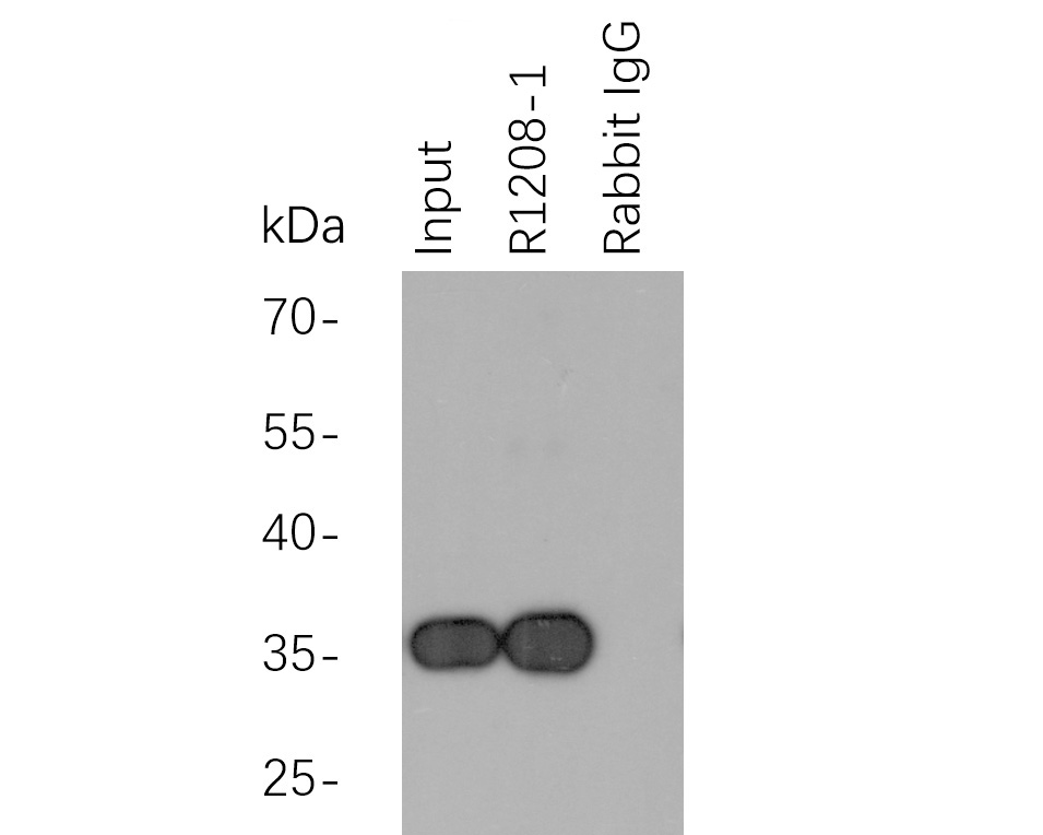
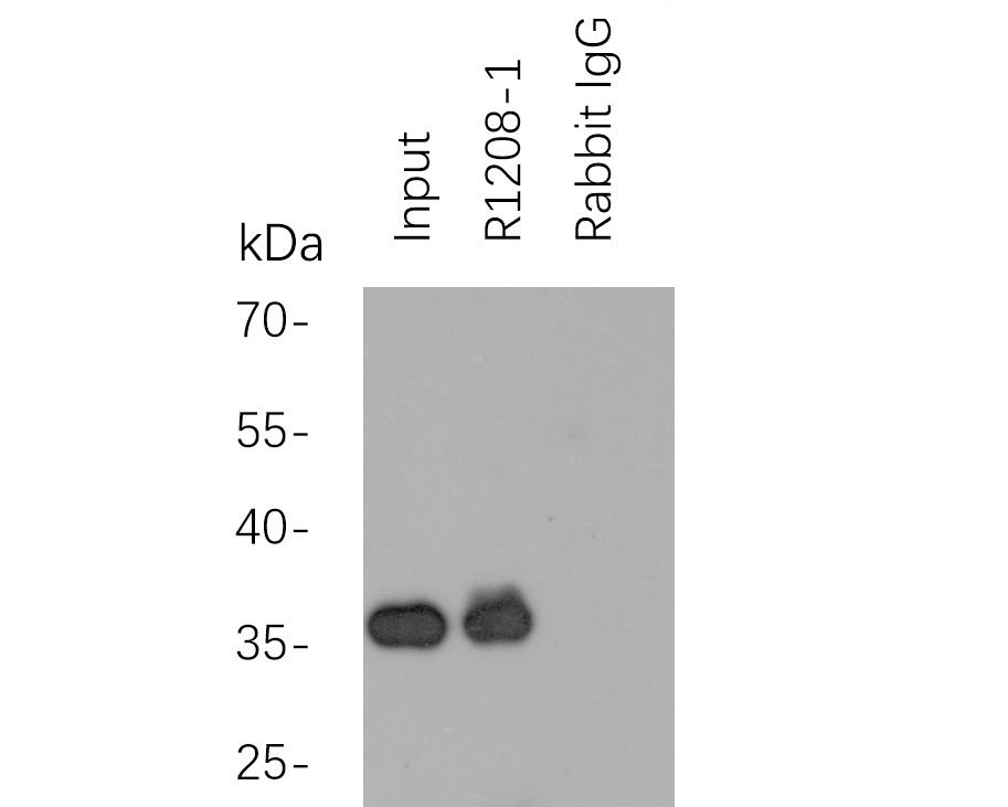
Myc tag was immunoprecipitated in 2µg C terminal Myc Tag fusion protein lysate with R1208-1 at 2 µg/20 µl agarose. Western blot was performed from the immunoprecipitate using EM31105 at 1/1000 dilution. Anti-Mouse IgG - HRP Secondary Antibody (HA1006) at 1:20,000 dilution was used for 60 mins at room temperature.
Lane 1: Myc Tag fusion protein lysate (input).
Lane 2: R1208-1 IP in Myc Tag fusion protein lysate.
Lane 3: Rabbit IgG instead of R1208-1 in Myc Tag fusion protein lysate.
Blocking/Dilution buffer: 5% NFDM/TBST

Myc tag was immunoprecipitated in 2µg N terminal Myc Tag fusion protein lysate with R1208-1 at 2 µg/20 µl agarose. Western blot was performed from the immunoprecipitate using EM31105 at 1/1000 dilution. Anti-Mouse IgG - HRP Secondary Antibody (HA1006) at 1:20,000 dilution was used for 60 mins at room temperature.
Lane 1: Myc Tag fusion protein lysate (input).
Lane 2: R1208-1 IP in Myc Tag fusion protein lysate.
Lane 3: Rabbit IgG instead of R1208-1 in Myc Tag fusion protein lysate.
Blocking/Dilution buffer: 5% NFDM/TBST

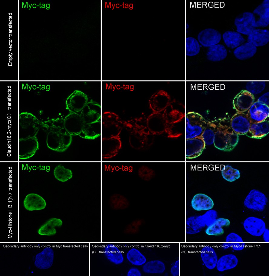
Immunocytochemistry analysis of 293T cells labeling Myc tag with Rabbit anti-Myc tag antibody (R1208-1) at 1/200 dilution.
293T cells, transfected with Myc-tagged empty control, Claudin18.2 (C-terminal) or Histone H3.1 (N-terminal) expression vector, respectively, were fixed in 4% paraformaldehyde for 10 minutes at room temperature, permeabilized with 0.1% Triton X-100 in PBS for 15 minutes at room temperature, then blocked with 1% BSA in 10% negative goat serum for 1 hour at room temperature. Cells were then incubated with Rabbit anti-Myc tag antibody (R1208-1) at 1/200 dilution in 1% BSA in PBST overnight at 4 ℃. Goat Anti-Rabbit IgG H&L (iFluor™ 594, HA1122) was used as the secondary antibody at 1/1,000 dilution. PBS instead of the primary antibody was used as the secondary antibody only control. Nuclear DNA was labelled in blue with DAPI.
Myc Tag (HA601081, green) was stained at 1/1,000 dilution overnight at +4℃. Goat Anti-Mouse IgG H&L (iFluor™ 488, HA1125) was used as the secondary antibody at 1/1,000 dilution.
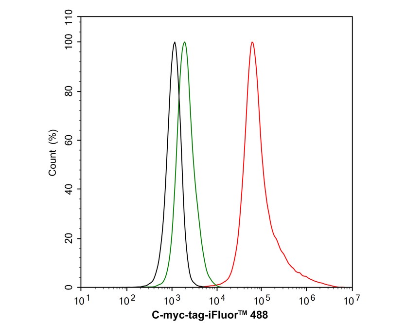
Flow cytometric analysis of 293T cells transfected C-myc-tag labeling Myc tag.
Cells were fixed and permeabilized. Then stained with the primary antibody (R1208-1, 1μg/mL) (red) compared with Rabbit IgG Isotype Control (green). After incubation of the primary antibody at +4℃ for an hour, the cells were stained with a iFluor™ 488 conjugate-Goat anti-Rabbit IgG Secondary antibody (HA1121) at 1/1,000 dilution for 30 minutes at +4℃. Unlabelled sample was used as a control (cells without incubation with primary antibody; black).

期刊: Veterinary Research
应用: WB,CoIP
反应种属: Human,Fish
发表时间: 2025 Feb


Huabio的大多数抗体是在甘油中提供的,所以抗体在推荐的-20℃储存条件下不会冻住。但是部分抗体未加甘油,我们建议进行分装使用,避免反复冻融。另外抗体分装可以减少操作不当引起的抗体污染带来的损失。


Copyright © 广州杰特伟生物科技有限公司 All Rights Reserved. 备案号:粤ICP备19077843号