HRP Conjugated Goat anti-Rat IgG(H+L) polyclonal Antibody
Goat Polyclonal Antibody
Rat IgG(H+L).
Rat
WB, ELISA
HRP
Liquid
2ug/ul
Store at +4℃ after thawing. Aliquot store at -20℃. Avoid repeated freeze / thaw cycles.
1*TBS (pH7.4), 0.5%BSA, 40% Glycerol.
IgG
Immunogen affinity purified.
WB
1:50,000-1:200,000
ELISA
1:5,000- 1:10,000
| WB | 查看 1 篇文献如下 |
| Dot Blot | 查看 1 篇文献如下 |
| Zebrafish | 查看 1 篇文献如下 |
| Mouse | 查看 1 篇文献如下 |
Whole IgG antibodies are isolated as intact molecules from antisera by immunoaffinity chromatography. They have an Fc portion and two antigen binding Fab portions joined together by disulfide bonds and therefore they are divalent. The average molecular weight is reported to be about 160 kDa. The whole IgG form of antibodies is suitable for the majority of immunodetection procedures and is the most cost effective. Horseradish peroxidase (HRP) conjugates are prepared by a modified Nakane and Kawaoi procedure (J. Histochem. Cytochem. 1974. 22, 1084). Peroxidase conjugates are commonly used for immunohistochemistry, Western blotting, and ELISA. Affinity-purified anti-horseradish peroxidase and conjugates are available for detection of horseradish peroxidase antigen or for signal amplification of HRP-containing reagents. For immunostaining of mammalian cells, an advantage of using anti-horseradish peroxidase is reduced background, since the antibody does not recognize the endogenous peroxidase-like enzymes found in those cells.
暂无
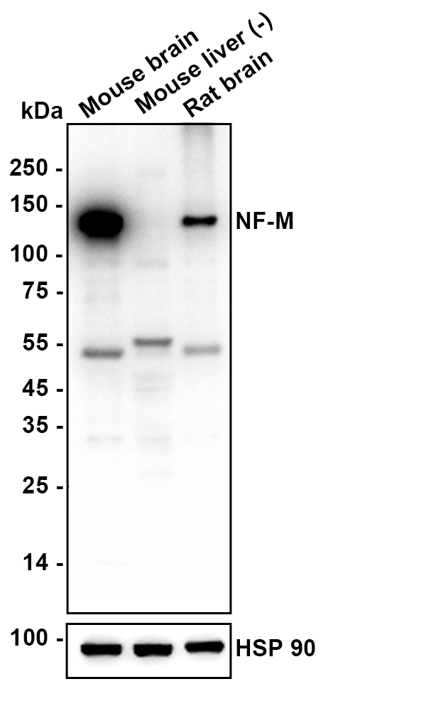
Western blot analysis of NF-M on different lysates with Rat anti-NF-M antibody (HA601408) at 1/5,000 dilution.
Lane 1: Mouse brain tissue lysate
Lane 2: Mouse liver tissue lysate (negative)
Lane 3: Rat brain tissue lysate
Lysates/proteins at 20 µg/Lane.
Predicted band size: 102 kDa
Observed band size: 140 kDa
Exposure time: 23 seconds; ECL: K1801;
4-20% SDS-PAGE gel.
Proteins were transferred to a PVDF membrane and blocked with 5% NFDM/TBST for 1 hour at room temperature. The primary antibody (HA601408) at 1/50,000 dilution was used in K1803 at 4℃ overnight. Goat Anti-Rat IgG - HRP Secondary Antibody (HA1023) at 1/50,000 dilution was used for 1 hour at room temperature.
CyHV-2 infection triggers mitochondrial-mediated apoptosis in GiCF cells by upregulating the pro-apoptotic gene ccBAX
Author: Cheng Wenjie,et al
PMID: 38253137
期刊: Fish & Shellfish Immunology
应用: WB
反应种属: Zebrafish
发表时间: 2024 Jan


Copyright © 广州杰特伟生物科技有限公司 All Rights Reserved. 备案号:粤ICP备19077843号